Retinal phototoxicity from the operating microscope is a photochemical injury of the macula after intraocular surgery.
It appears as a small, oval lesion near the fovea within a day or two.
Therefore, it is worth knowing when a patient has unexplained poor vision after an uneventful operation.
What Happens in Retinal Phototoxicity
The intense light of the microscope focuses on the macula through a clear cornea and lens.
Short wavelengths are the most harmful and cause photochemical damage.
Also, the damage is not thermal and does not need high power.
Because the fovea sits in the focal path, it is the usual site.
Historical Description
McDonald and Irvine described light-induced maculopathy after extracapsular cataract extraction in 1983.
Later studies in phacoemulsification confirmed the problem.
Also, Byrnes and colleagues followed patients prospectively after surgery.
Therefore, the entity is well documented.
Fundus Explorer Pro
Photograph the retinal findings described here with the phone already in your pocket — 22 D optics and built-in illumination in one handheld unit.
From Choroida — the team behind this siteRisk Factors for Retinal Phototoxicity
Long operating time is the main factor.
Light intensity, coaxial illumination and a fixed focus add to the risk.
Also, clear media and lighter fundus pigmentation increase exposure.
Because even short cases can cause lesions, avoid assuming safety.
Clinical Picture of Retinal Phototoxicity
Patients report blurred vision, a central scotoma or a color change.
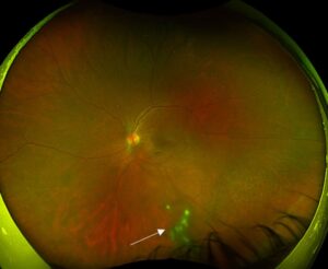
The fundus shows a yellow-white lesion at or near the fovea in the first days.
Also, the lesion later turns into a pigmented or atrophic scar.
Because the findings are subtle, a careful macular examination is required.
Differential Diagnosis of Retinal Phototoxicity
Postoperative cystoid macular edema causes blurred vision weeks after surgery (see Irvine-Gass syndrome).
Solar retinopathy produces a similar lesion from sun gazing (see solar retinopathy).
Also, laser pointer injury causes a related picture (see laser pointer maculopathy).
Therefore, the history of surgery guides the diagnosis.
Imaging Findings
Optical coherence tomography shows disruption of the ellipsoid zone and the outer retina.
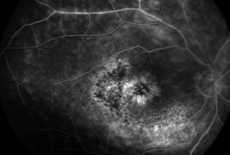
Fluorescein angiography may show a window defect.
Also, the changes may be small and need careful review (see ellipsoid zone disruption).
Because the lesion is tiny, use high-resolution scans.
Natural Course of Retinal Phototoxicity
Vision may improve partly over weeks to months.
Some patients keep a small central defect.
Also, the outcome depends on the depth of injury.
Therefore, counsel patients with a realistic message.
Preventing Retinal Phototoxicity During Surgery
Reduce the light intensity to the minimum that allows a good view.
Use filters that block short wavelengths when possible.
Also, shift the beam off the macula with a light shield or by tilting the microscope.
Because exposure adds up, shorten the time of full illumination.
Endoilluminators in Vitrectomy
The light pipe also causes phototoxicity during vitrectomy.
Keep the tip away from the macula and use low intensity.
Also, avoid fixed illumination over the fovea.
Therefore, apply the same principles as in cataract surgery.
Medicolegal Aspects
Photic injury is a known complication and needs documentation.
Record the duration of light exposure and the steps taken to reduce it.
Also, tell the patient about the diagnosis in plain terms.
Because trust matters, be open and supportive.
Practical Summary
Think of phototoxicity in a patient with poor central vision after clear surgery.
Examine the macula and obtain OCT.
Also, reduce light exposure in every case.
Wavelengths and Light Safety
Blue and near-ultraviolet light cause the most photochemical damage.
Longer wavelengths are less harmful but can cause heat.
Also, standards set limits for light exposure during surgery.
Because filters help, microscope manufacturers include them.
Risk in Pseudophakic and Aphakic Eyes
The natural lens filters short wavelengths.
After the lens is removed, the retina is more exposed.
Also, an intraocular lens without a filter may transmit more blue light.
Therefore, take extra care in the last stage of cataract surgery.
Symptoms and Visual Outcome
Patients may notice a central scotoma and mild blur.
Some recover completely, and others keep a small defect.
Also, visual acuity depends on foveal involvement.
Because the course varies, follow the patient for several months.
Other Light-Related Retinal Injuries
Welding arc injury and exposure to bright light cause similar changes.
Also, prolonged exposure to a fiber optic light during retinal surgery can do so.
Therefore, ask about all sources of intense light.
Operative Notes and Safety Culture
Record the intensity settings and the approximate duration of exposure.
Also, review the technique in teaching sessions.
Because awareness reduces injury, share cases within the team.
For the latest studies, see the PubMed results for retinal phototoxicity.


Document what you see
Two smartphone imaging tools built for everyday clinic use — one for the slit lamp, one for the fundus.
From Choroida — the team behind this siteReferences
- Michels M, Sternberg P Jr. Operating microscope-induced retinal phototoxicity: pathophysiology, clinical manifestations and prevention. Surv Ophthalmol. 1990;34:237-252.
- McDonald HR, Irvine AR. Light-induced maculopathy from the operating microscope in extracapsular cataract extraction and intraocular lens implantation. Ophthalmology. 1983;90:945-951.
- Byrnes GA, Antoszyk AN, Mazur DO, Kao TC, Miller SA. Photic maculopathy after extracapsular cataract surgery: a prospective study. Ophthalmology. 1992;99:731-738.
- Wu J, Seregard S, Algvere PV. Photochemical damage of the retina. Surv Ophthalmol. 2006;51:461-481.

Writes and reviews the clinical articles on Educate. Every post is checked against peer-reviewed sources before it goes live.