Scleral lens complications are uncommon when the lens is well fitted, but they can threaten the cornea if they are missed.
These lenses vault over the cornea and rest on the sclera, and they hold a reservoir of fluid on the surface.
That design brings benefits for irregular and diseased corneas, and it brings its own problems (see scleral lens fitting).
Why Scleral Lens Complications Arise
- The fluid reservoir and the lens reduce the oxygen that reaches the cornea
- The lens edge rests on the conjunctiva and sclera and can also compress it
- Debris and mucus collect in the fluid under the lens
- The lens is large, so handling and cleaning are more demanding
- Patients often have severely diseased eyes, which are more vulnerable
Scleral Lens Complications: Hypoxia and Corneal Edema
Poor oxygen reaches the cornea if the lens is thick, the material has a low oxygen permeability, or the central vault is high.
- Symptoms: blurred vision after removal, haloes, and discomfort at the end of the day
- Signs: corneal edema, microcysts, and later neovascularization (see contact lens hypoxia)
- Prevention: use a high-permeability material, a thin lens, and the lowest vault that clears the cornea, and limit the hours of wear
All-fit Slit-Lamp Adapter
Record and share exactly what you see at the slit lamp. One adapter fits any slit lamp or surgical microscope — and any smartphone.
From Choroida — the team behind this siteMidday Fogging
Many patients notice a gradual clouding of vision over the day, caused by debris, lipids, and cells that drift into the fluid reservoir.
- It is more frequent in patients with meibomian gland disease and in those with high vault
- Management includes removing and reapplying the lens with fresh solution, treating the lid disease, reducing the vault, and using a different solution or lens surface
Debris may also contain inflammatory cells, so persistent fogging needs attention.
Scleral Lens Complications: Conjunctival Problems
- Impingement: the edge digs into the conjunctiva and may also cause blanching, a ring-shaped depression, and redness
- Prolapse and staining of the conjunctiva
- Limbal compression, with reduced circulation
- Conjunctivochalasis, which can interfere with the lens edge (see conjunctivochalasis)
The remedy is a change in the landing zone, with a flatter or steeper edge, or a toric or quadrant-specific design.
Infection and Inflammation
Scleral lenses can cause microbial keratitis, a rare and serious complication, especially in eyes with a damaged surface (see contact lens microbial keratitis risk factors).
- Use sterile preservative-free saline, never tap water, to fill the lens
- Teach the cleaning routine, and warn about the risk of contaminated cases
- Stop wear and review at once for pain or redness
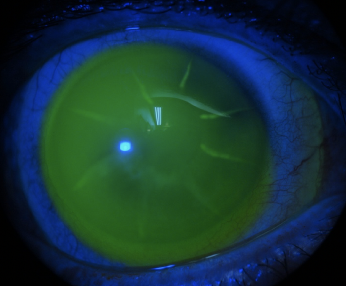
Clinicians also see sterile infiltrates and giant papillary conjunctivitis.
Other Problems
- Suction and lens adherence: the lens sticks to the eye and is hard to remove, which can be avoided by a better edge design and by teaching a proper technique
- Intraocular pressure rise: from compression of the sclera in some eyes, especially those with glaucoma (see normal tension glaucoma)
- Bubbles under the lens, from poor filling
- Endothelial changes with long-term wear, which should be monitored with specular microscopy
Follow-Up for Scleral Lens Complications
- Review at one to two weeks after the first fit, and then every three to six months
- Check the vault, the landing zone, the corneal clarity, and the conjunctiva
- Use anterior segment OCT, where it is available, to measure the vault and the edge
- Check the pressure in eyes with glaucoma
Regular review identifies problems before they cause damage.
Counselling
Explain the care routine, the importance of hygiene, and the signs that need an urgent visit.
Reinforce that the lens should be removed at the first sign of pain or redness.
Handling and Cleaning Tips
- Fill the lens bowl with preservative-free saline, and avoid bubbles
- Apply and remove with clean hands and the correct tool, such as a small plunger
- Clean with a recommended solution, and rinse before storage
- Replace the case every few months, and use fresh solution each day
Mistakes in handling are a common cause of problems, and a short demonstration at each visit pays off.
When to Refit
Consider a refit if there is persistent fogging, impingement, edema, or discomfort, or after a major change in the eye, such as a corneal graft or cross-linking.
Use a new measurement of the shape of the sclera and cornea, and anterior segment OCT, if available, to guide the change.
Small adjustments to the vault, the edge, or the material may also resolve most problems.
Keep a log of each patient’s lens parameters, vault, and landing zone findings, so that small changes are easy to see over time and can guide the next adjustment.
For the latest studies, see the PubMed results for scleral lens complications.


Document what you see
Two smartphone imaging tools built for everyday clinic use — one for the slit lamp, one for the fundus.
From Choroida — the team behind this siteReferences
- Visser ES, Visser R, van Lier HJ, Otten HM. Modern scleral lenses part I: clinical features. Eye Contact Lens. 2007;33:13-20.
- Pecego M, Barnett M, Mannis MJ, Durbin-Johnson B. Jupiter scleral lenses: the UC Davis Eye Center experience. Eye Contact Lens. 2012;38:179-182.
- Michaud L, van der Worp E, Brazeau D, Warde R, Giasson CJ. Predicting estimates of oxygen transmissibility for scleral lenses. Cont Lens Anterior Eye. 2012;35:266-271.
- American Academy of Ophthalmology. Basic and Clinical Science Course, Section 8: External Disease and Cornea. AAO; current edition.

Writes and reviews the clinical articles on Educate. Every post is checked against peer-reviewed sources before it goes live.