
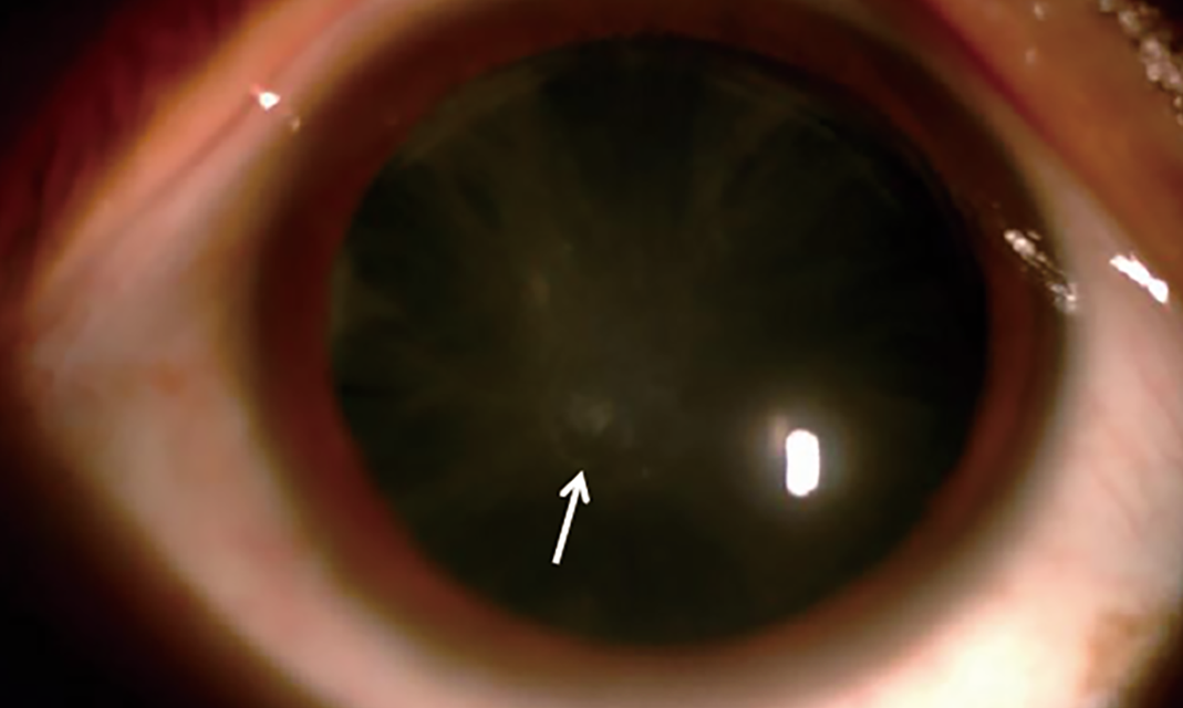
A patient comes in with a few small, sparkling flecks in the lens, and the examiner wonders whether the lens is merely aging.
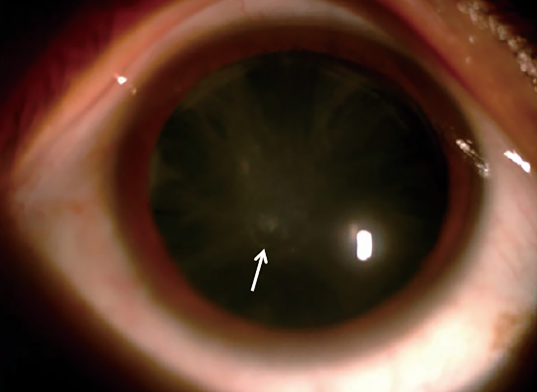
When the opacities are fine, crystalline, and located in the cortex just beneath the capsule, in a person with a history of neck surgery or tetany, it is worth thinking about hypocalcemic cataract.
The underlying problem
Low serum calcium that persists over months or years, usually because of hypoparathyroidism or pseudohypoparathyroidism, interferes with the permeability of lens cell membranes.
The lens depends on calcium to keep its membrane channels intact, and chronic hypocalcemia disturbs the ion balance and allows protein aggregation to occur.
The most common cause is accidental removal or injury to the parathyroid glands during thyroid or neck surgery.
Idiopathic hypoparathyroidism, autoimmune polyglandular disease, and genetic syndromes such as DiGeorge are other causes.
Malabsorption, vitamin D deficiency, and chronic kidney disease can also produce chronic hypocalcemia.
What it looks like
- Numerous small, discrete white or iridescent dots in the anterior and posterior subcapsular cortex
- A clear zone between the opacities and the capsule, which gives the lens a characteristic layered appearance
- Sparing of the nucleus in early disease
- Progression, in a proportion of patients, to dense subcapsular and cortical opacity
Bilateral involvement is typical, and the findings may be symmetrical.
All-fit Slit-Lamp Adapter
Record and share exactly what you see at the slit lamp. One adapter fits any slit lamp or surgical microscope — and any smartphone.
From Choroida — the team behind this siteOther signs of hypocalcemia
The eye is only one part of the story.
Patients may have perioral numbness, muscle cramps, carpopedal spasm, positive Chvostek and Trousseau signs, prolonged QT interval, and calcification of the basal ganglia.
Papilledema from raised intracranial pressure can also occur in hypoparathyroidism.
In children, delayed dentition and poor growth may be seen.
Investigations
- Serum calcium (corrected for albumin), phosphate, magnesium, and parathyroid hormone
- 25-hydroxyvitamin D and renal function
- Electrocardiogram
- Neuroimaging when neurological signs are present
- Genetic testing if a syndromic cause is suspected
Treatment
The treatment is medical, with calcium and active vitamin D analogues, and adequate magnesium, titrated to keep calcium in the low normal range.
Once calcium is normalized, the progression of lens changes often stops, and in some young patients fine opacities may stabilize.
Established dense cataracts do not reverse.
When the cataract affects vision, cataract surgery is performed in the usual way.
Calcium should be stable before surgery to avoid perioperative problems such as tetany, and the endocrinologist should be involved.
What the lens looks like in practice
Early on, the flecks are tiny and glitter like crystals under the slit lamp, sitting in the cortex just inside the capsule. They tend to be most obvious with retroillumination and a dilated pupil. The nucleus is usually clear. If hypocalcemia is corrected early, the flecks may remain unchanged for years, and in a few young patients they seem to fade.
Interpreting the finding in a patient with neck surgery
A history of thyroidectomy or parathyroidectomy, even decades earlier, is a clue. Ask about tingling, cramps, and the use of calcium supplements. If the patient is not on treatment, check the calcium and parathyroid hormone, and refer to an endocrinologist. If the patient is on treatment, ask whether the levels have been kept in range, and send a short letter to the treating physician about the lens findings.
Other eye findings
Hypoparathyroidism may be associated with papilledema, optic neuritis, blepharospasm, and conjunctival calcification. A full eye examination, including the disc, is worthwhile. Patients with long-standing low calcium should be reviewed every year or two, particularly children, who can develop dense cataract over a short time.
Surgery
When cataract surgery is needed, the surgeon should confirm normal calcium levels in the preceding days. Corneal and anterior segment findings are usually routine, and the visual result is generally good.
How to work with the endocrinologist
Share the lens findings in a letter, and ask for the current calcium, phosphate, and parathyroid hormone values, along with the treatment plan. Mention that eye findings can progress if control is poor, and ask whether the patient has other complications such as kidney stones or basal ganglia calcification. Joint care often leads to better control and prevents unnecessary surgery.
Children and adolescents
In children, hypocalcemia may be a manifestation of DiGeorge syndrome or of autoimmune polyendocrine disease. A child with cataract and seizures, tetany, or developmental problems needs urgent pediatric evaluation. Early treatment of the underlying disease may prevent further lens damage.
Differential diagnosis
Other causes of small cortical opacities include steroid use, diabetes, myotonic dystrophy, and Wilson disease. The pattern of fine iridescent flecks, together with a low serum calcium, is characteristic of the hypocalcemic form.
Prognosis
With good control of calcium, the prognosis for the lens is favorable.
Poorly controlled hypoparathyroidism, particularly in a child or adolescent, can lead to progressive cataract that needs surgery at a young age.


Document what you see
Two smartphone imaging tools built for everyday clinic use — one for the slit lamp, one for the fundus.
From Choroida — the team behind this siteReferences
- Shoback D. Hypoparathyroidism. N Engl J Med. 2008;359:391-403.
- Bronsky D, Kushner DS, Dubin A, Snapper I. Idiopathic hypoparathyroidism and pseudohypoparathyroidism; case reports and review of the literature. Medicine. 1958;37:317-352.
- Mitchell DM, Regan S, Cooley MR, et al. Long-term follow-up of patients with hypoparathyroidism. J Clin Endocrinol Metab. 2012;97:4507-4514.
- Brandi ML, Bilezikian JP, Shoback D, et al. Management of hypoparathyroidism: summary statement and guidelines. J Clin Endocrinol Metab. 2016;101:2273-2283.
