Conjunctivochalasis is a common, often underrecognized cause of chronic tearing and ocular irritation, in which loose, redundant folds of bulbar conjunctiva, typically along the inferior lid margin, mechanically interfere with normal tear drainage and tear film stability, producing symptoms that are frequently mistaken for simple dry eye or nasolacrimal duct obstruction until the conjunctiva itself is examined closely.
What Conjunctivochalasis Is
Conjunctivochalasis results from a loss of the normal firm attachment between the bulbar conjunctiva and the underlying sclera, related to age-related degeneration of the connective tissue anchoring the conjunctiva, allowing the conjunctiva to become loose and redundant, most typically along the inferior fornix and lid margin.
Why Redundant Conjunctiva Causes Tearing
This is one of the more counterintuitive aspects of the condition, since patients with conjunctivochalasis present with tearing, a symptom more often associated with too much fluid rather than a surface fold.
- The redundant conjunctival tissue physically disrupts the tear meniscus along the lower lid, interfering with the normal tear film reservoir and its distribution across the ocular surface
- The loose folds can mechanically obstruct the punctum, the tear drainage opening, particularly during blinking, impairing normal tear outflow
- Redundant conjunctiva can undergo mechanical irritation with blinking, producing a secondary reflex tearing response
- The combination of disrupted tear film distribution and impaired drainage produces both an unstable tear film (with dry eye-like surface symptoms) and overflow tearing (epiphora), sometimes in the same patient
All-fit Slit-Lamp Adapter
Record and share exactly what you see at the slit lamp. One adapter fits any slit lamp or surgical microscope — and any smartphone.
From Choroida — the team behind this siteClinical Presentation
- Chronic tearing (epiphora), often the presenting complaint, sometimes leading to an initial, incorrect workup for nasolacrimal duct obstruction
- Ocular irritation, foreign body sensation, and mild discomfort, particularly with blinking
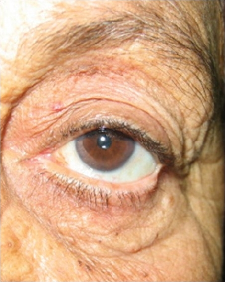
- Visible loose, redundant folds of conjunctiva, most prominent along the inferior bulbar conjunctiva near the lid margin, often more apparent on downgaze or with gentle lid manipulation during examination
- Symptoms and findings are often more prominent temporally or nasally depending on the pattern of conjunctival redundancy
- A relatively normal-appearing tear film on basic testing in some patients, which can make the diagnosis easy to overlook if the conjunctiva itself is not specifically examined
Diagnosis
Diagnosis is clinical, based on careful slit-lamp examination looking specifically for redundant conjunctival folds along the lower lid margin, particularly when a patient presents with tearing that has not responded to standard dry eye or punctal evaluation.
Excluding other causes of epiphora, including nasolacrimal duct obstruction (which can be assessed with lacrimal irrigation) and punctal stenosis, is an important part of the workup, since conjunctivochalasis can coexist with, or be mistaken for, these other causes of tearing.
Management
Conservative Measures
- Lubrication with artificial tears, which can provide some symptomatic relief, though it does not address the underlying mechanical cause
- Topical anti-inflammatory treatment for any associated surface inflammation
Surgical Management
For patients with significant, persistent symptoms not adequately relieved by conservative measures, surgical excision of the redundant conjunctival tissue is the definitive treatment.
- Conjunctival resection, removing the excess, redundant conjunctiva and allowing the remaining tissue to heal in a tauter, better-anchored position, is the standard surgical approach
- Various techniques exist for managing the resulting conjunctival defect, including primary closure, allowing healing by secondary intention, or using adjunctive materials such as amniotic membrane in more extensive cases
- Thermal or radiofrequency cautery techniques have also been described as alternative or adjunctive approaches to reduce conjunctival redundancy
Outcomes
Surgical treatment of conjunctivochalasis generally provides good relief of both the tearing and irritation symptoms in appropriately selected patients, restoring more normal tear meniscus formation and punctal apposition.
Careful patient selection, confirming that conjunctivochalasis is genuinely the primary driver of symptoms rather than a coexisting but non-causative finding, supports the best surgical outcomes, since redundant conjunctiva is common with advancing age and not all cases are symptomatic enough to warrant surgical intervention.
Clinical Significance
Recognizing conjunctivochalasis as a distinct, specifically treatable cause of chronic tearing, separate from the more commonly considered causes such as dry eye and nasolacrimal duct obstruction, prevents both unnecessary and ineffective treatment directed at the wrong underlying problem and, when surgery is appropriate, offers a good chance of definitive symptom relief.


Document what you see
Two smartphone imaging tools built for everyday clinic use — one for the slit lamp, one for the fundus.
From Choroida — the team behind this siteReferences
- Meller D, Tseng SC. Conjunctivochalasis: literature review and possible pathophysiology. Surv Ophthalmol. 1998;43:225-232.
- Di Pascuale MA, Espana EM, Kawakita T, Tseng SC. Clinical characteristics of conjunctivochalasis with or without aqueous tear deficiency. Br J Ophthalmol. 2004;88:388-392.
- Youm DJ, Kim JM, Choi CY. Simple surgical approach with high-frequency radio-wave electrosurgery for conjunctivochalasis. Ophthalmology. 2010;117:2129-2133.
- Balci O. Clinical evaluation of conjunctivochalasis with and without dry eye. Ocul Surf. 2014;12:65-71.
Test yourself
A few questions straight from this article.
-
What defines conjunctivochalasis?
Conjunctivochalasis is loose, redundant bulbar conjunctiva, typically along the inferior lid margin, which mechanically interferes with tear drainage and tear film stability. -
What underlies the conjunctival redundancy in conjunctivochalasis?
The firm attachment between bulbar conjunctiva and underlying sclera is lost through age-related degeneration of the anchoring connective tissue, letting the conjunctiva become loose and redundant. -
How do redundant conjunctival folds impair tear outflow in conjunctivochalasis?
The loose folds can physically block the punctum, especially during blinking, impairing normal outflow and contributing to overflow tearing. -
Which tear film structure is disrupted by the folds in conjunctivochalasis?
Redundant tissue physically disrupts the lower lid tear meniscus, interfering with the tear film reservoir and its distribution across the ocular surface. -
Which presenting complaint in conjunctivochalasis often triggers the wrong initial workup?
Chronic epiphora is often the presenting complaint and can lead to an initial, incorrect workup for nasolacrimal duct obstruction before the conjunctiva itself is examined. -
What makes the conjunctival folds of conjunctivochalasis more apparent at the slit lamp?
The redundant folds along the inferior bulbar conjunctiva are often more obvious on downgaze or with gentle lid manipulation, which is why the diagnosis is missed when the conjunctiva is not specifically examined. -
Which test helps exclude nasolacrimal duct obstruction in a patient with suspected conjunctivochalasis?
Nasolacrimal duct obstruction can be assessed with lacrimal irrigation, and excluding it along with punctal stenosis is part of the workup because these causes can coexist with or mimic conjunctivochalasis. -
What is the limitation of artificial tears in conjunctivochalasis?
Lubrication can give some symptomatic relief but does nothing about the underlying mechanical problem of redundant conjunctiva obstructing drainage and disrupting the meniscus. -
What is the definitive treatment for symptomatic conjunctivochalasis?
Conjunctival resection, removing the excess tissue so the remainder heals in a tauter, better-anchored position, is the standard definitive operation when conservative measures fail. -
What most supports a good surgical outcome in conjunctivochalasis?
Redundant conjunctiva is common with advancing age and not always symptomatic, so confirming it genuinely drives the symptoms rather than being a coexisting finding underpins the best results.

Writes and reviews the clinical articles on Educate. Every post is checked against peer-reviewed sources before it goes live.