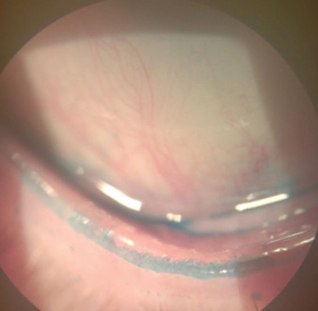
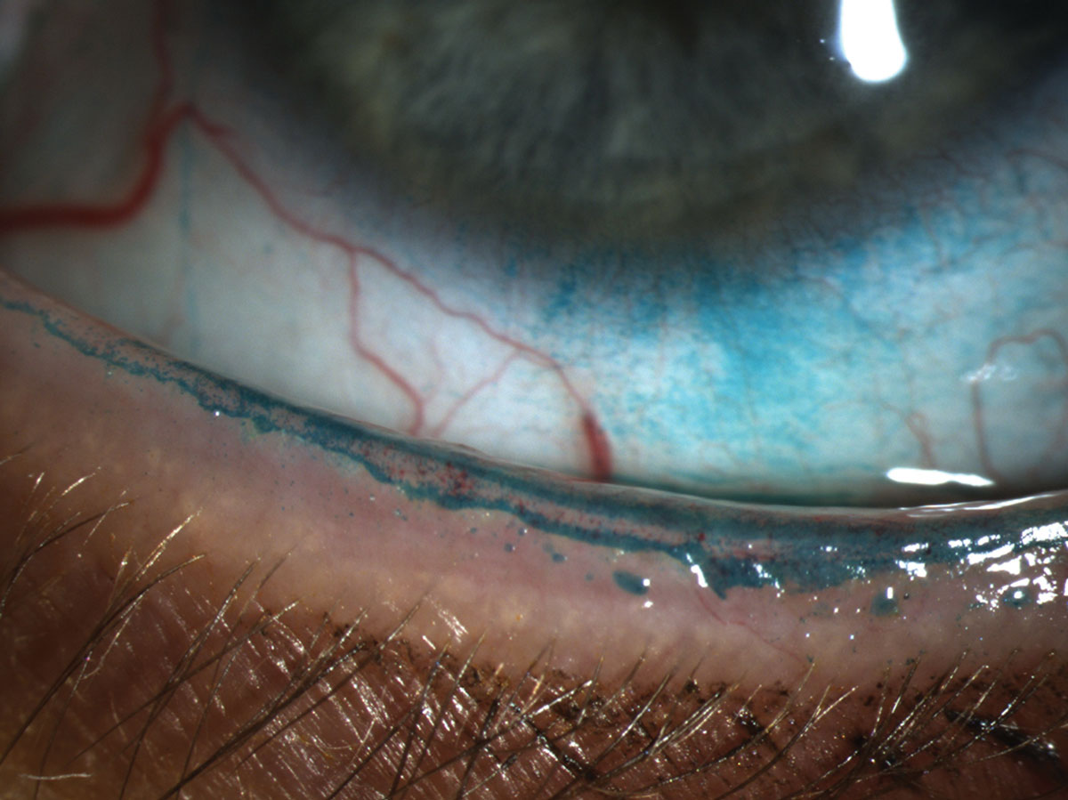
The Marx line is a line along the margin of the eyelid, seen when the lid is stained with lissamine green or fluorescein.
It marks the border between the wet surface of the conjunctiva and the dry skin of the lid, and its position carries information about the health of the meibomian glands.
It takes a few seconds to look for, but it is often forgotten.
What Is the Marx Line?
The lid margin has a skin portion in front, the gland orifices in the middle, and the conjunctival portion behind.
Also, staining the lid margin shows a thin line of dye at the mucocutaneous junction, where the dye clings to the tissue.
Marx described this line, and later studies suggested that it can also be used as a marker of the lid margin condition (see the references).
Marx Line: Normal Position
In a healthy lid, the line lies behind the meibomian gland orifices, on the conjunctival side, and is thin and regular.
The orifices are also open, and the oil flows over the lid margin to form the tear film lipid layer.
All-fit Slit-Lamp Adapter
Record and share exactly what you see at the slit lamp. One adapter fits any slit lamp or surgical microscope — and any smartphone.
From Choroida — the team behind this siteWhat Changes in Disease
- In meibomian gland dysfunction, the line moves forward, to or in front of the gland orifices
- The line becomes thick, irregular, or broken
- It may cross the orifices, so that the orifices appear to lie within the stained area
- In severe disease, the lid margin shows keratinization and notches
Studies show that the line shifts with the severity of gland dysfunction, and a forward shift correlates with more severe changes (see the references).
Marx Line: How to Look for It
- First, use a drop of fluorescein or a lissamine green strip, applied to the lower lid or the conjunctiva
- Then ask the patient to blink a few times to spread the dye
- Examine the upper and lower lid margins at the slit lamp, with low magnification and then higher
- Use a cobalt blue filter for fluorescein, and white light for lissamine green
- Finally, evert the upper lid to see the upper margin
Check the position of the line relative to the orifices, and its width and regularity.
Related Lid Margin Findings
- Lid wiper epitheliopathy: staining on the inner lid margin where it wipes the cornea, linked to friction and dryness (see lid wiper epitheliopathy)
- Telangiectasia and thickening of the margin
- Plugged orifices and foamy secretions (see meibomian gland dysfunction)
- Gland dropout on imaging (see meibography)
These findings, together, build a complete picture of the lid margin.
Why It Matters
- It gives a quick, cheap clue to gland disease in a patient with dry eye symptoms
- It may also explain symptoms in patients with a normal tear break-up time
- Also, it helps to monitor the response to lid treatment
- It provides a simple way to teach students and patients
However, it is not a diagnostic test on its own, and its use varies among clinicians.
Marx Line: Management When the Line Is Abnormal
- Treat the lid margin disease with warm compresses, lid massage, and hygiene
- Consider thermal pulsation or intense pulsed light in selected patients
- Use antibiotic or anti-inflammatory drugs where there is inflammation (see blepharitis)
- Treat associated dry eye, and also advise on lubricants
Review at intervals to see whether the line returns to a normal position.
A Quick Routine for the Lid Margin
- Look at the lid margin and lashes with the slit lamp before instilling any drops
- Press gently on the lower lid to express the glands, and note the secretion
- Stain with lissamine green or fluorescein, and check the position of the line and any staining of the margin
- Evert the upper lid and examine the margin and the conjunctiva
This takes about two minutes and adds helpful information to the dry eye evaluation.
Reporting and Follow-Up
Record whether the line lies behind, at, or in front of the orifices, and whether it is regular or broken.
Compare findings after treatment, and show the patient the change.
Limits of Interpretation
There is variation in how clinicians identify the line, while results depend on the stain and the technique.
The lid margin changes with age, and some degree of forward movement is common in older patients, so interpret the finding with the rest of the examination.
Also, combine it with the findings of gland expressibility, tear stability, and corneal staining to form a complete picture (see dry eye disease).
For the latest studies, see the PubMed results for Marx line.


Document what you see
Two smartphone imaging tools built for everyday clinic use — one for the slit lamp, one for the fundus.
From Choroida — the team behind this siteReferences
- Yamaguchi M, Kutsuna M, Uno T, et al. Marx line: fluorescein staining line on the inner lid as an indicator of meibomian gland function. Am J Ophthalmol. 2006;141:669-675.
- Korb DR, Blackie CA. Meibomian gland diagnostic expressibility: correlation with dry eye symptoms and gland location. Cornea. 2008;27:1142-1147.
- Korb DR, Herman JP, Blackie CA, et al. Prevalence of lid wiper epitheliopathy in subjects with dry eye signs and symptoms. Cornea. 2010;29:377-383.
- American Academy of Ophthalmology. Basic and Clinical Science Course, Section 8: External Disease and Cornea. AAO; current edition.

Writes and reviews the clinical articles on Educate. Every post is checked against peer-reviewed sources before it goes live.