
Few complaints are as common in an eye clinic — and as easy to under-treat — as “my eyes feel dry and irritated.”
Dry eye disease (DED) is not a single diagnosis but a spectrum of tear film and ocular surface dysfunction.
It ranges from mild, intermittent irritation to a chronic condition that meaningfully erodes quality of life and visual function.
Symptoms and clinical signs frequently disagree with each other, which is part of why DED is so often mismanaged.
A patient can have significant surface disease with few symptoms, or severe symptoms with a relatively unremarkable exam.
Understanding where a given patient sits on the aqueous-deficient to evaporative spectrum is what actually determines whether treatment works.
What Is Dry Eye Disease?
Dry eye disease (DED) is a multifactorial disease of the ocular surface characterized by a loss of tear film homeostasis, accompanied by ocular symptoms in which tear film instability and hyperosmolarity, ocular surface inflammation and damage, and neurosensory abnormalities play etiological roles.
Per the TFOS DEWS II framework, DED is classified along a spectrum:
- Aqueous-deficient dry eye (ADDE) — insufficient tear production by the lacrimal gland
- Evaporative dry eye (EDE) — excessive tear evaporation, most often from meibomian gland dysfunction, but also blink- or ocular surface-related
- Mixed disease — most patients show features of both subtypes rather than a pure form
These subtypes are best understood as points on a single spectrum of disease rather than as entirely separate conditions.
Epidemiology
DED is one of the most common conditions encountered in eye care.
- Symptomatic prevalence estimates range roughly from 5% to 50%, depending on the population and diagnostic criteria used
- Prevalence based on clinical signs alone is generally higher and more variable, reaching up to 75% in some populations
- Prevalence increases with age, with signs increasing more steeply per decade than symptoms
- Women have a consistently higher prevalence than men, particularly from midlife onward
The wide range in reported prevalence reflects a real clinical challenge: signs and symptoms of DED frequently do not correlate well with each other.
All-fit Slit-Lamp Adapter
Record and share exactly what you see at the slit lamp. One adapter fits any slit lamp or surgical microscope — and any smartphone.
From Choroida — the team behind this sitePathophysiology
Regardless of the initiating subtype, DED converges on a shared, self-perpetuating cycle.
- Reduced aqueous tear production or increased evaporation raises tear film osmolarity
- Hyperosmolar tears trigger inflammatory signaling at the ocular surface
- Inflammation damages the corneal and conjunctival epithelium and injures goblet cells that produce mucin
- Mucin loss and epithelial damage further destabilize the tear film
- This instability accelerates evaporation and re-elevates osmolarity, perpetuating the cycle
Neurosensory changes at the ocular surface also contribute, which is why symptom severity does not always track cleanly with the degree of visible surface damage.
Risk Factors
Ocular Risk Factors
- Meibomian gland dysfunction and blepharitis
- Contact lens wear
- Prior ocular surgery, particularly refractive surgery that can affect corneal nerve sensitivity
- Reduced blink rate from prolonged screen use
Systemic and Demographic Risk Factors
- Increasing age
- Female sex, particularly postmenopausal
- Autoimmune and connective tissue disease, especially Sjögren syndrome
- Certain systemic medications, including antihistamines, some antidepressants, and isotretinoin
- Vitamin A deficiency and other nutritional factors, in specific populations
A quick review of medications and any autoimmune history is often more diagnostically useful than the eye exam alone.
Clinical Presentation
Symptoms
- Burning, stinging, or a gritty foreign-body sensation
- Fluctuating, mildly blurred vision that often improves transiently with blinking
- Paradoxical watering, from reflex tearing in response to surface irritation
- Symptoms typically worse later in the day, in low-humidity environments, or with prolonged screen use
Examination Findings
- Reduced tear meniscus height
- Rapid or irregular tear film break-up on the ocular surface
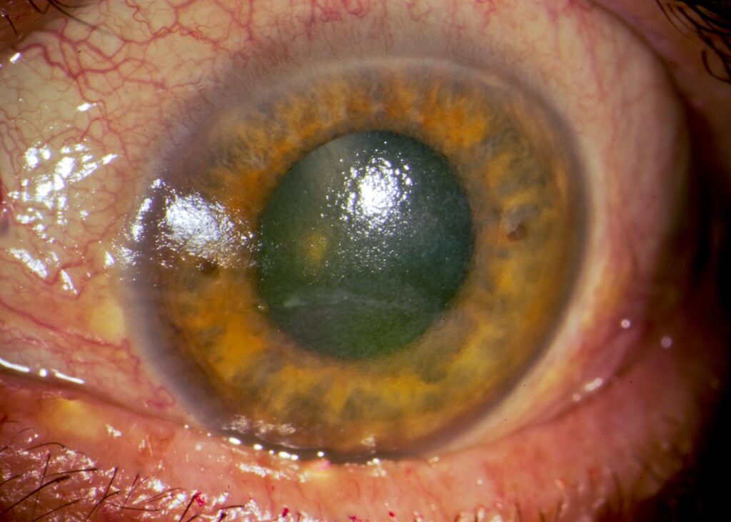
- An irregular, dulled corneal light reflex reflecting surface irregularity
- Punctate epithelial erosions on the cornea, often more pronounced in the interpalpebral zone
- Conjunctival hyperemia and, in more severe cases, filaments or mucus strands
Clinical signs and reported symptom severity frequently diverge, which is exactly why relying on either alone risks under- or over-treating the patient in front of you.
Diagnostic Evaluation
Symptom Assessment
- Validated questionnaires (e.g., OSDI, SPEED) provide a structured, reproducible symptom score and a way to track response to treatment
Tear Film Testing
- Tear break-up time assesses tear film stability
- Tear osmolarity testing, where available, directly measures the hyperosmolarity central to disease pathophysiology
- Schirmer testing evaluates aqueous tear production, useful when aqueous deficiency is suspected
Ocular Surface Staining
- Fluorescein staining highlights corneal epithelial disruption and quantifies punctate erosions
- Lissamine green or rose bengal staining highlights conjunctival and devitalized epithelial cells, particularly useful in milder disease
Meibomian Gland Assessment
- Gland expression evaluates secretion quality and patency
- Meibography, where available, images gland structure and dropout directly
Combining a symptom score with at least one objective sign-based test is the most reliable way to confirm the diagnosis and classify its subtype.
Differential Diagnosis
Conditions that can mimic or coexist with DED include:
- Blepharitis and meibomian gland dysfunction — frequently a contributing cause rather than a true mimic
- Allergic conjunctivitis — itching predominates, often with a seasonal or exposure-related pattern
- Ocular surface neuropathic pain — disproportionate symptoms relative to minimal clinical signs
- Sjögren syndrome — severe aqueous-deficient dry eye with systemic sicca symptoms and positive autoantibodies
- Contact lens-related dryness and discomfort — closely tied to wear time and lens type
- Filamentary keratitis — a more severe presentation with mucus filaments adherent to the corneal surface
Screening for an underlying autoimmune cause matters most in younger patients or those with disproportionately severe aqueous deficiency.
Management
First-Line Measures
- Preservative-free artificial tears, tailored to whether the disease is more aqueous-deficient or evaporative in nature
- Warm compresses and lid hygiene when meibomian gland dysfunction contributes
- Environmental modification — humidifiers, reduced screen time, and scheduled blinking during prolonged near work
Anti-Inflammatory and Prescription Therapy
- Topical cyclosporine or lifitegrast for chronic, inflammation-driven disease not controlled by tears alone
- Short courses of topical corticosteroids for flares of surface inflammation, used judiciously given long-term risks
- Oral omega-3 fatty acid supplementation, though evidence for benefit is mixed across trials
Procedural and Advanced Options
- Punctal plugs to conserve the natural tear film in predominantly aqueous-deficient disease
- In-office thermal pulsation or intense pulsed light therapy for refractory meibomian gland dysfunction
- Scleral lenses or autologous serum tears for severe, treatment-resistant ocular surface disease
Treatment should be matched to the dominant subtype and severity rather than escalating through a generic ladder for every patient.
Prognosis
DED is a chronic condition managed rather than cured.
- Most patients achieve good symptom control with consistent first-line and, where needed, anti-inflammatory therapy
- Severity tends to fluctuate with environmental factors, screen use, and systemic disease activity
- Untreated, longstanding disease can progress to corneal surface complications, including persistent epithelial defects or, rarely, infectious keratitis
Consistency of therapy, not intensity of a single visit’s treatment, is what determines long-term outcomes.
Would you like to document anterior segment findings with your smartphone?


Document what you see
Two smartphone imaging tools built for everyday clinic use — one for the slit lamp, one for the fundus.
From Choroida — the team behind this siteReferences
- Craig JP, Nichols KK, Akpek EK, et al. TFOS DEWS II Definition and Classification Report. The Ocular Surface. 2017.
- Stapleton F, Alves M, Bunya VY, et al. TFOS DEWS II Epidemiology Report. The Ocular Surface. 2017.
- Bron AJ, de Paiva CS, Chauhan SK, et al. TFOS DEWS II Pathophysiology Report. The Ocular Surface. 2017.
- Jones L, Downie LE, Korb D, et al. TFOS DEWS II Management and Therapy Report. The Ocular Surface. 2017.
- American Academy of Ophthalmology. Dry Eye Syndrome Preferred Practice Pattern.
Test yourself
A few questions straight from this article.
-
Dry eye disease is defined by the loss of which central homeostatic feature?
Dry eye disease is a multifactorial ocular surface disease characterised by loss of tear film homeostasis, with tear instability, hyperosmolarity, inflammation and neurosensory abnormality all playing a role. -
Under the TFOS DEWS II framework, dry eye disease is classified along which spectrum?
TFOS DEWS II places dry eye on a spectrum from aqueous-deficient to evaporative disease, with most patients showing mixed features rather than a pure subtype. -
What is the reported range of symptom-based prevalence estimates for dry eye disease?
Symptomatic prevalence estimates range from about 5% to 50% depending on population and diagnostic criteria, while sign-based prevalence is higher and reaches up to 75% in some populations. -
What triggers inflammatory signalling in the self-perpetuating cycle of dry eye disease?
Reduced tear production or increased evaporation raises tear osmolarity, and hyperosmolar tears trigger surface inflammation that damages epithelium and goblet cells, further destabilising the tear film. -
Where are punctate epithelial erosions typically most pronounced in dry eye disease?
Fluorescein-positive punctate epithelial erosions in dry eye are often most marked in the interpalpebral zone, the strip of cornea exposed between the lids. -
Which test directly measures the hyperosmolarity central to dry eye pathophysiology?
Tear osmolarity testing measures hyperosmolarity directly, whereas break-up time assesses tear film stability and Schirmer testing assesses aqueous production. -
Symptoms far out of proportion to minimal clinical signs should raise which alternative diagnosis?
Ocular surface neuropathic pain classically produces disproportionate symptoms alongside minimal objective signs, and is an important mimic of dry eye disease. -
What is the first-line pharmacological measure for dry eye disease?
Preservative-free artificial tears, chosen according to whether the disease is more aqueous-deficient or evaporative, are the first-line measure alongside lid hygiene and environmental modification. -
Which topical prescription therapies target chronic inflammation-driven dry eye disease?
Topical cyclosporine or lifitegrast is used for chronic inflammation-driven dry eye that is not controlled by artificial tears alone, with short steroid courses reserved for flares. -
Which corneal complication can follow untreated, longstanding dry eye disease?
Longstanding untreated disease can progress to corneal surface complications, including persistent epithelial defects and, rarely, infectious keratitis.

Writes and reviews the clinical articles on Educate. Every post is checked against peer-reviewed sources before it goes live.


