
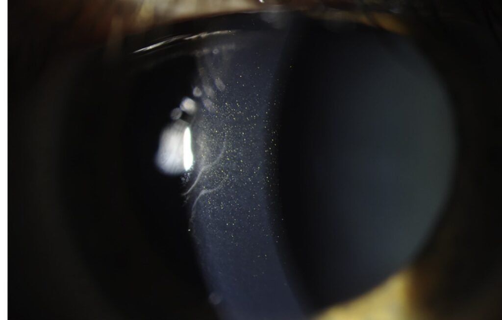
Pre-Descemet corneal dystrophy is a group of mostly benign conditions in which small opacities lie in the deepest layers of the stroma, just in front of Descemet membrane.
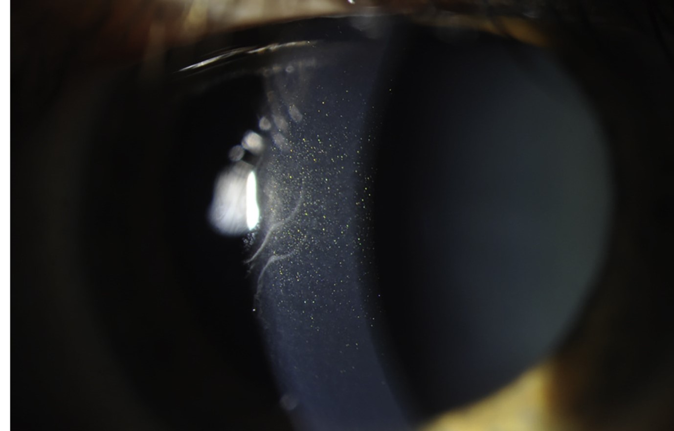
They are found in adults, usually after the age of 30, and most patients have no visual symptoms.
The main clinical task is to recognize them as a variant and to avoid a work-up for a disease that is not there.
What Is Pre-Descemet Corneal Dystrophy?
The opacities are fine, gray-white, and often polymorphic, and they may take the shape of dots, commas, filaments, boomerangs, or short curved lines.
They are seen best with a narrow beam and retroillumination, and they lie directly anterior to the endothelium and Descemet membrane.
The central and peripheral cornea are both affected, and the overlying stroma and epithelium are normal.
Cornea farinata is a closely related pattern, in which the dots are so small that they look like flour dusted on the posterior stroma.
Some cases are familial, with autosomal dominant inheritance, and others appear to be sporadic or related to age.
Isolated and Secondary Forms
The classification used today separates the dystrophy from a larger group of secondary pre-Descemet changes.
A pre-Descemet pattern has been reported with X-linked ichthyosis, in which steroid sulfatase deficiency produces deep stromal opacities in many patients.
Similar findings occur with several systemic and metabolic conditions, and with some drugs.
Because of this, a family history, a skin examination, and a review of medications are worth a few minutes when the pattern is unusually dense or appears in a young patient.
In an older patient with a typical bilateral pattern and good vision, no further investigation is needed.
All-fit Slit-Lamp Adapter
Record and share exactly what you see at the slit lamp. One adapter fits any slit lamp or surgical microscope — and any smartphone.
From Choroida — the team behind this siteClinical Examination
Use a slit lamp with a moderate beam width and look at the deep stroma at an angle.
Scanning the cornea in slices helps to see the full extent of the opacities and to confirm that they are located in front of Descemet membrane.
The endothelial mosaic should be examined in specular reflection, because it should be regular in this condition.
Corneal thickness, intraocular pressure, and sensation are normal.
Photographs made at the first visit are useful for comparison, because the pattern is stable over many years.
Differential Diagnosis
- Fleck corneal dystrophy: discrete flat gray flecks throughout the stroma (see fleck corneal dystrophy)
- Fuchs endothelial dystrophy: guttae and thickening of Descemet membrane, with edema as the disease advances (see Fuchs endothelial corneal dystrophy)
- Posterior polymorphous dystrophy: vesicles and bands at the level of Descemet membrane with endothelial changes (see posterior polymorphous corneal dystrophy)
- Posterior crocodile shagreen: polygonal opacities with clear spaces between them
- Drug deposits: chloroquine, amiodarone, and other agents produce epithelial or subepithelial deposits more often than deep stromal ones
Management
No treatment is needed when the visual acuity is good, and the opacities do not progress in a way that changes this.
Patients can be told that the finding is benign, that it is not related to the early stage of Fuchs dystrophy, and that routine follow-up is enough.
In a rare case with unusually dense opacities or reduced vision, a different diagnosis should be considered first.
If the patient is a boy or young man with dry, scaly skin, a diagnosis of ichthyosis can be raised with the pediatrician or dermatologist.
Why the Distinction Matters in Practice
The main risk of mislabeling a deep stromal opacity is to attach the wrong prognosis to it.
A patient told that they have an early endothelial dystrophy may undergo unnecessary monitoring and anxiety, whereas a patient with a true endothelial dystrophy may miss follow-up if the finding is dismissed as an age change.
Specular microscopy and pachymetry settle the question quickly, because the endothelial cell mosaic and central thickness are normal in pre-Descemet dystrophy and abnormal in Fuchs dystrophy.
Imaging
Anterior segment OCT shows the opacities as small hyperreflective foci in the deep stroma, with a normal Descemet membrane line behind them.
In vivo confocal microscopy can show highly reflective bodies in the posterior stroma and may reveal that the keratocyte nuclei look normal.
These tests are optional, and they are most useful when the clinician is documenting a case for a family or for teaching.
Counselling
Explain that the opacities are located in a layer that does not take part in normal vision, and that they do not usually change with time.
Mention that the finding may be discovered again by another examiner in the future, and a short note in the chart avoids repeated work-ups.
If a family member is found to have a similar pattern, it can be taken as supporting a familial form, and no treatment is required for either of them.


Document what you see
Two smartphone imaging tools built for everyday clinic use — one for the slit lamp, one for the fundus.
From Choroida — the team behind this siteReferences
- Weiss JS, Møller HU, Aldave AJ, et al. IC3D classification of corneal dystrophies, edition 2. Cornea. 2015;34:117-159.
- Mannis MJ, Holland EJ, eds. Cornea. 4th ed. Elsevier; 2017.
- Salmon JF. Kanski’s Clinical Ophthalmology: A Systematic Approach. 9th ed. Elsevier; 2020.
- American Academy of Ophthalmology. Basic and Clinical Science Course, Section 8: External Disease and Cornea. AAO; current edition.

Writes and reviews the clinical articles on Educate. Every post is checked against peer-reviewed sources before it goes live.


