
CASE REPORT
A 45-year-old female presented with persistent eye discomfort, foreign body sensation, burning, itching, occasional blurry vision, and worsening symptoms during reading or computer use. Previous use of over-the-counter lubricating eye drops provided no relief.
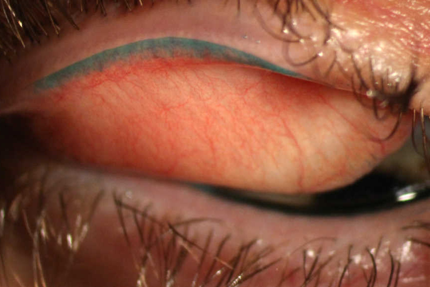
Clinical examination showed normal visual acuity, unremarkable anterior segment, but redness and increased vascularity in the lid wiper region of both upper eyelids. Clear corneas ruled out corneal involvement, while decreased tear breakup time indicated poor tear film stability.
Diagnostic tests, including Schirmer’s test and fluorescein staining, supported the diagnosis of Lid-wiper epitheliopathy.
Lid-wiper epitheliopathy DISEASE entity
Lid-wiper epitheliopathy is defined as a disruption to the surface epithelium of the lid-wiper, a portion of the marginal conjunctiva of the upper and lower lid that acts as a wiping surface to spread the tear film over the ocular surface. It is often observed as staining of the lid wiper area by vital dyes.
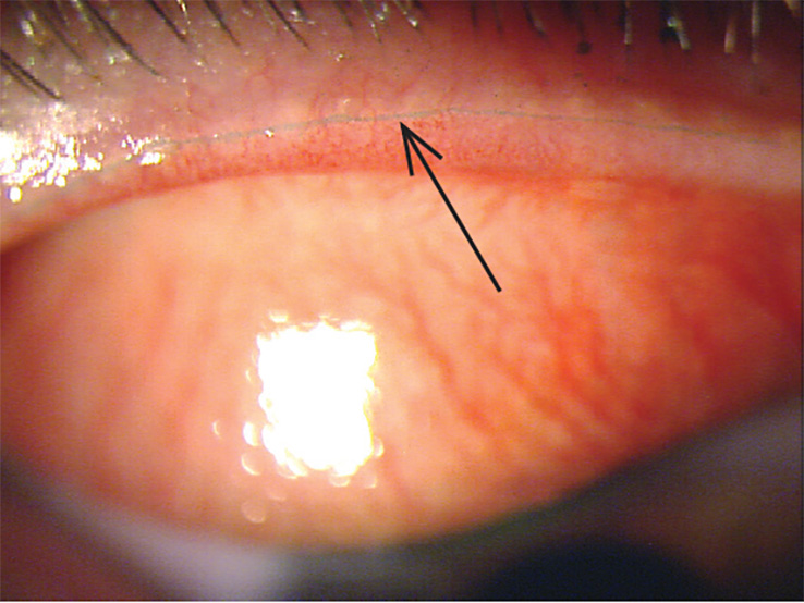
The etiology of Lid-wiper epitheliopathy is likely multifactorial, with different causes proposed for those associated with contact lens wear and those associated with dry eye symptoms without contact lens wear.
One of the leading hypotheses is increased friction between the lid wiper and the ocular or anterior contact lens surface, leading to physical trauma and mechanical abrasion of the epithelial cells of the lid wiper. Hyperosmotic insult from changes in tear osmolarity may play an important role in lower LWE as the lower lid wiper is less subject to friction-related damage due to a shorter excursion distance during blinks.
There may also be an inflammatory component as upregulation of inflammatory cells has been observed. Due to the association between Lid-wiper epitheliopathy and dry eye symptoms, many risk factors of LWE overlap with those of dry eye, including but not limited to:
- Contact lens wear (both soft and rigid), especially in those with symptoms of dry eye.
- Presence of dry eye symptoms in non-contact lens wearers
- Older age
- Asian race
- Low humidity and increased airflow environments
Activities that provoke front surface drying and/or incomplete/infrequent blinking, such as prolonged reading and/or computer use.
All-fit Slit-Lamp Adapter
Record and share exactly what you see at the slit lamp. One adapter fits any slit lamp or surgical microscope — and any smartphone.
From Choroida — the team behind this siteLid-wiper epitheliopathy Diagnosis
Lid-wiper epitheliopathy is primarily diagnosed by observation of lid-wiper staining by vital dyes, the defining feature of the condition. Because of its common association with dry eye symptoms and/or contact lens wear, a detailed history of dry eye-related symptoms and contact lens-associated discomfort should be collected to differentiate and/or rule out other similar conditions when establishing the diagnosis.
MANAGEMENT of Lid-wiper epitheliopathy
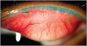
Current therapies for Lid-wiper epitheliopathy have been studied either by directly assessing their benefits on LWE or using LWE as a surrogate marker in the course of alleviating dry eye or contact lens discomfort.
Medical therapy
Several options have demonstrated efficacy in treating Lid-wiper epitheliopathy and its associated signs and symptoms in preliminary studies, including topical corticosteroids, oil-in-water emulsion lubricant eye drops, basic fibroblast growth factors, and topical rebamipide.
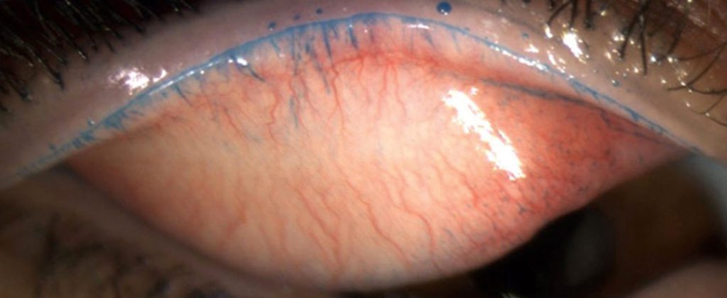
Insertion of punctal plugs alleviated signs and symptoms of aqueous tear deficient dry eye, including Lid-wiper epitheliopathy. Other treatments that have been proposed include strategies to improve blinking behavior, which may be beneficial in LWE associated with incomplete blinking behaviors, as well as improving contact lens wear comfort. Topical vitamin A ointment may be helpful.
The efficacy of these strategies remains to be validated further.


Document what you see
Two smartphone imaging tools built for everyday clinic use — one for the slit lamp, one for the fundus.
From Choroida — the team behind this siteREFERENCES
- Korb DR, Greiner JV, Herman JP, et al. Lid-wiper epitheliopathy and dry-eye symptoms in contact lens wearers. Eye & Contact Lens. 2002;28(4):211–16.
- Efron N, Brennan NA, Morgan PB, Wilson T. Lid wiper epitheliopathy. Progress in Retinal and Eye Research. 2016;53:140-174. doi: 10.1016/j.preteyeres.2016.04.004.
- Korb DR, Herman JP, Greiner JV, et al. Lid wiper epitheliopathy and dry eye symptoms. Eye and Contact Lens. 2005;31(1):2-8. Accessed Nov 28, 2020. doi: 10.1097/01.ICL.0000140910.03095.FA.
- McMonnies CW. An examination of the relationship between ocular surface tear osmolarity compartments and epitheliopathy. Ocular Surface. 2015;13(2):110-117. Accessed Nov 28, 2020. doi: 10.1016/j.jtos.2014.07.002.
- Golebiowski B, Chim K, So J, Jalbert I. Lid margins: Sensitivity, staining, meibomian gland dysfunction, and symptoms. Optometry and Vision Science. 2012;89(10):1443–1449. doi: 10.1097/OPX.0b013e3182693cef.
- Morgan P, Petropoulos I, Read M, Malik R, Maldonado-Codina C. Confocal microscopy of the lid margin area of contact lens wearers. Contact Lens and Anterior Eye. 2013;36:e43.
Test yourself
A few questions straight from this article.
-
Lid-wiper epitheliopathy is defined as disruption of the epithelium of which structure?
The lid wiper is the portion of marginal conjunctiva on the upper and lower lid that spreads the tear film across the ocular surface; epitheliopathy is disruption of its surface epithelium. -
Which is the leading proposed mechanism for lid-wiper epitheliopathy?
Friction between the lid wiper and the ocular or anterior contact lens surface is thought to cause physical trauma and mechanical abrasion of lid-wiper epithelial cells. -
Why is hyperosmotic insult thought to matter more for lower lid-wiper epitheliopathy?
Because the lower lid wiper travels a shorter distance during blinks, it sustains less friction-related damage, so changes in tear osmolarity are proposed to play a larger role there. -
What is the defining diagnostic feature of lid-wiper epitheliopathy?
Diagnosis rests primarily on observing lid-wiper staining with vital dyes, supported by a detailed history of dry eye symptoms and contact lens discomfort. -
Which environmental exposure is listed as a risk factor for lid-wiper epitheliopathy?
Risk factors overlap with those of dry eye and include low humidity and increased airflow environments, alongside contact lens wear, older age and Asian race. -
Which behaviour pattern is implicated in provoking lid-wiper epitheliopathy?
Activities that provoke front surface drying and incomplete or infrequent blinking, such as prolonged reading and computer work, are implicated in lid-wiper epitheliopathy. -
Which slit lamp finding was present in this case of lid-wiper epitheliopathy?
The anterior segment was otherwise unremarkable with clear corneas; the abnormality was redness and increased vascularity in the lid-wiper region of both upper eyelids. -
Which tear film abnormality supported the diagnosis in this case of lid-wiper epitheliopathy?
A decreased tear breakup time indicated poor tear film stability, and Schirmer testing with fluorescein staining supported the diagnosis of lid-wiper epitheliopathy. -
Which topical agents have shown preliminary efficacy in lid-wiper epitheliopathy?
Preliminary studies support topical corticosteroids, oil-in-water emulsion lubricant drops, basic fibroblast growth factors and topical rebamipide, though efficacy still needs validation. -
Which intervention relieved lid-wiper epitheliopathy in aqueous tear deficient dry eye?
Punctal plugs alleviated the signs and symptoms of aqueous tear deficient dry eye, including lid-wiper epitheliopathy; improving blinking behaviour has also been proposed.

Writes and reviews the clinical articles on Educate. Every post is checked against peer-reviewed sources before it goes live.


