
Meibomian gland dysfunction is chronic abnormality of the meibomian glands, most often obstruction of their orifices and thickening of the oil they produce, and it is now recognized as the leading cause of evaporative dry eye disease.
That distinction, evaporative rather than aqueous-deficient dry eye, matters directly for treatment, since artificial tears alone address the wrong side of the tear film when the actual problem is an unstable, rapidly evaporating oil layer.
Meibomian gland dysfunction is extremely common, and it coexists with or drives a meaningful share of the dry eye and blepharitis cases seen in routine practice.
Prevalence rises with age, and by later adulthood some degree of meibomian gland dysfunction is present in a substantial majority of people, even when it never becomes symptomatic enough to prompt an eye exam.
Normal Meibomian Gland Function
The meibomian glands, embedded in the tarsal plates of both eyelids, secrete meibum, an oily substance that forms the outermost layer of the tear film and slows evaporation of the aqueous layer beneath it.
Normal meibum is clear and flows easily with gentle expression, spreading evenly across the tear film with each blink to maintain a stable, slow-evaporating tear surface.
When gland function is disrupted, either by obstruction of the orifices or qualitative changes in the secretion itself, the tear film’s evaporative barrier fails, and symptoms of ocular surface irritation follow.
Pathophysiology
Obstructive meibomian gland dysfunction, the most common form, involves hyperkeratinization and plugging of the gland orifices, causing meibum to thicken, stagnate, and eventually become difficult to express even with manual pressure.
Over time, chronic obstruction can lead to gland dropout, a permanent loss of functioning glandular tissue visible on meibography as areas of gland atrophy that do not recover even once obstruction is treated.
Demodex mite infestation of the lash follicles and glands, androgen deficiency, and chronic low-grade inflammation of the lid margin all contribute to the obstructive process in different patients, which is part of why treatment often needs to be individualized rather than applying a single protocol to everyone.
Rosacea and other inflammatory skin conditions are commonly associated as well, and a patient’s facial skin findings can offer a useful clue to the underlying driver of their meibomian gland disease.
All-fit Slit-Lamp Adapter
Record and share exactly what you see at the slit lamp. One adapter fits any slit lamp or surgical microscope — and any smartphone.
From Choroida — the team behind this siteClinical Presentation
- Burning, foreign body sensation, and fluctuating blurred vision, often worse later in the day as tear film instability accumulates over hours of open-eye activity
- Symptoms that seem disproportionate to how mild the eye looks on casual inspection, a common source of patient frustration when prior exams have not specifically assessed gland function
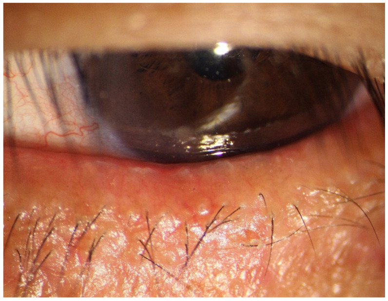
- Capped or plugged meibomian gland orifices visible on careful slit-lamp exam of the lid margin, sometimes with surrounding telangiectasia
- A rapid tear break-up time on fluorescein staining, reflecting the unstable, evaporation-prone tear film typical of significant meibomian gland dysfunction
Evaluation
Diagnosis is largely clinical, based on lid margin examination, gland expressibility, and the quality of expressed meibum, ranging from clear and easily expressed to thick, toothpaste-like, or completely inspissated.
Tear break-up time and, where available, meibography, which images the glands directly to assess structural dropout, add objective documentation and help gauge how much of the problem is reversible obstruction versus permanent gland loss.
Distinguishing meibomian gland dysfunction from aqueous-deficient dry eye, and recognizing that many patients have elements of both, shapes which treatments are likely to help most.
Management
Warm compresses and lid hygiene remain first-line treatment, softening thickened meibum and mechanically clearing obstructed orifices to restore more normal gland outflow.
In-office thermal expression devices provide a more standardized, sustained heat and pressure treatment than compresses alone can reliably achieve, and can be useful for patients who have not responded adequately to home care.
Oral tetracycline-class antibiotics, used for their anti-inflammatory rather than antimicrobial effect, and topical azithromycin are both used to reduce the chronic lid margin inflammation that perpetuates obstruction in many patients.
Artificial tears and other tear film supplements remain useful adjuncts for symptom relief, but they treat the downstream tear film instability rather than the underlying glandular problem, which is why they alone rarely resolve significant meibomian gland dysfunction.
Because gland dropout seen on meibography does not reverse, setting realistic expectations early matters: the goal in advanced disease is stabilizing symptoms and preserving remaining gland function, not restoring glands that have already atrophied.


Document what you see
Two smartphone imaging tools built for everyday clinic use — one for the slit lamp, one for the fundus.
From Choroida — the team behind this siteReferences
- Nichols KK, Foulks GN, Bron AJ, et al. The International Workshop on Meibomian Gland Dysfunction: executive summary. Investigative Ophthalmology & Visual Science.
- American Academy of Ophthalmology. Basic and Clinical Science Course, Section 8: External Disease and Cornea.
- Craig JP, Nichols KK, Akpek EK, et al. TFOS DEWS II definition and classification report. The Ocular Surface.
Test yourself
A few questions straight from this article.
-
Meibomian gland dysfunction is recognised as the leading cause of which problem?
Chronic meibomian gland abnormality, usually orifice obstruction with thickened oil, is the leading cause of evaporative dry eye, which is a different problem from aqueous deficiency. -
What does meibum contribute to the tear film?
Meibum from glands in the tarsal plates forms the outermost tear film layer and slows evaporation of the aqueous layer beneath it. -
What characterises the obstructive form of meibomian gland dysfunction?
The commonest form involves hyperkeratinization and plugging of orifices, so meibum thickens, stagnates and becomes hard to express even with manual pressure. -
What does gland dropout on meibography signify in meibomian gland dysfunction?
Chronic obstruction can cause gland dropout, permanent atrophy visible on meibography that does not recover even once the obstruction is treated. -
Which skin condition is commonly associated with meibomian gland dysfunction?
Rosacea and other inflammatory skin conditions are commonly associated, so a patient's facial skin findings can point to the driver of their gland disease. -
Which fluorescein finding reflects the unstable tear film of significant meibomian gland dysfunction?
Tear break-up time is rapid because the deficient oil layer leaves an unstable, evaporation-prone tear film. -
When are the symptoms of meibomian gland dysfunction typically worst during the day?
Burning, foreign body sensation and fluctuating blur are often worse later in the day as instability accumulates over hours of open-eye activity. -
What remains first-line treatment for meibomian gland dysfunction?
Warm compresses and lid hygiene soften thickened meibum and mechanically clear obstructed orifices, restoring more normal gland outflow. -
Why are oral tetracycline-class antibiotics used in meibomian gland dysfunction?
Tetracyclines, like topical azithromycin, are used to reduce the chronic lid margin inflammation that perpetuates obstruction, not for an antibacterial effect. -
Why do artificial tears alone rarely resolve significant meibomian gland dysfunction?
Tear supplements usefully relieve symptoms but address the resulting tear film instability rather than the obstructed, dysfunctional glands driving it.

Writes and reviews the clinical articles on Educate. Every post is checked against peer-reviewed sources before it goes live.


