Chronic eye irritation that never quite goes away often has a simple, overlooked source: the eyelid margin itself.
Blepharitis is inflammation of the lid margin, and it is one of the most common conditions seen in any eye clinic.
It rarely threatens vision directly, but it drives a disproportionate share of chronic ocular surface complaints.
Patients often cycle through artificial tears and antibiotic drops without lasting relief because the underlying lid margin disease is never specifically addressed.
Blepharitis is not a single disease — it spans several distinct mechanisms, from bacterial colonization to meibomian gland dysfunction to Demodex mite infestation.
Correctly classifying which type (or combination) a patient has is what actually determines whether treatment works.
What Is Blepharitis?
Blepharitis is a chronic inflammatory condition of the eyelid margin, broadly divided by anatomic location.
The two principal forms are:
- Anterior blepharitis — affects the eyelid skin, lash follicles, and lash base; further subdivided into staphylococcal and seborrheic types
- Posterior blepharitis — affects the meibomian glands, and largely overlaps with meibomian gland dysfunction (MGD)
Many patients have mixed anterior and posterior disease, and Demodex mite infestation can contribute to either form.
Epidemiology
Blepharitis is extremely common and likely underreported, since many patients simply live with mild symptoms.
- It is among the most frequent reasons for referral to eye care for ocular surface complaints
- Prevalence increases with age, and posterior blepharitis/MGD becomes proportionally more common in older adults
- Demodex folliculorum and Demodex brevis mite colonization increases markedly with age and is found in a substantial proportion of adults over 60, though colonization alone does not equal disease
Because blepharitis is chronic and relapsing rather than curable, most patients require ongoing lid hygiene rather than a single course of treatment.
All-fit Slit-Lamp Adapter
Record and share exactly what you see at the slit lamp. One adapter fits any slit lamp or surgical microscope — and any smartphone.
From Choroida — the team behind this sitePathophysiology
The mechanism differs meaningfully between the two anatomic forms.
- Staphylococcal anterior blepharitis involves chronic bacterial colonization of the lash follicles, provoking a low-grade inflammatory and sometimes toxin-mediated reaction
- Seborrheic anterior blepharitis is associated with excess sebaceous gland secretion and is often linked to seborrheic dermatitis elsewhere on the skin
- Posterior blepharitis/MGD results from obstruction and qualitative change in meibomian gland secretions, which destabilizes the tear film lipid layer
- Demodex folliculorum preferentially colonizes the lash base, contributing to anterior disease, while Demodex brevis inhabits the meibomian glands, contributing to posterior disease
Whichever mechanism dominates, the end result is the same: an unstable tear film, chronic surface inflammation, and recurrent symptoms.
Risk Factors
Ocular Risk Factors
- Pre-existing dry eye disease or tear film instability
- Rosacea-associated ocular surface disease
- Contact lens wear
- History of chalazia, which frequently coexist with untreated MGD
Demographic and Systemic Risk Factors
- Increasing age
- Seborrheic dermatitis
- Diabetes, which is associated with a higher burden of MGD in some studies
Because most risk factors are chronic rather than acute, blepharitis management is realistically about long-term control, not a one-time cure.
Clinical Presentation
Symptoms
- Burning, itching, or a foreign-body sensation, typically worse in the morning
- Eyelid crusting on waking, sometimes with lashes matted together
- Redness of the lid margins
- Fluctuating, mildly blurred vision related to an unstable tear film
- Chronic, relapsing course with symptom-free intervals
Examination Findings
Findings vary by subtype:
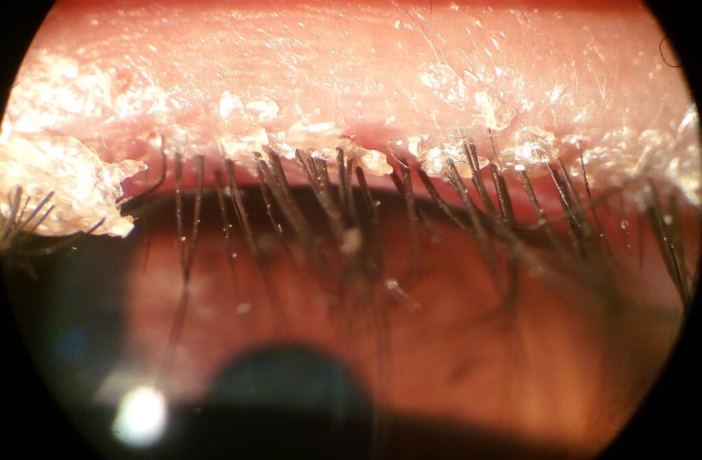
- Cylindrical collarettes wrapped around the lash base — characteristic of Demodex infestation
- Hard, brittle crusts and occasional lash loss or misdirection — typical of staphylococcal blepharitis
- Greasy, scaly debris along the lid margin — typical of seborrheic blepharitis
- Thickened, irregular lid margins with plugged or capped meibomian gland orifices — typical of posterior blepharitis/MGD
- Reduced or poor-quality meibum expression on gentle lid pressure
Careful slit-lamp inspection of the lash base and gland orifices — not just the general appearance of redness — is what actually distinguishes the subtypes.
Diagnostic Evaluation
Slit-Lamp Examination
- Direct inspection of lash base, lid margin, and meibomian gland orifices remains the primary diagnostic tool
- Epilation of a lash for microscopic examination can confirm Demodex when collarettes are seen but the diagnosis is uncertain
Tear Film Assessment
- Tear break-up time is often reduced given the frequent overlap with evaporative dry eye
- Meibomian gland expression assesses secretion quality and gland patency
No laboratory test is routinely required — blepharitis remains a clinical diagnosis made at the slit lamp.
Differential Diagnosis
Conditions that can mimic or coexist with blepharitis include:
- Dry eye disease — often coexists with and is worsened by blepharitis, rather than being a true mimic
- Chalazion/hordeolum — focal nodules rather than diffuse lid margin change
- Allergic conjunctivitis — itching predominates, with conjunctival rather than lid margin findings
- Herpes simplex or zoster blepharitis — vesicular lesions and a more acute, often unilateral presentation
- Sebaceous gland carcinoma — must be considered in unilateral, treatment-resistant, or atypical chronic blepharitis, particularly in older patients
Distinguishing routine blepharitis from these mimics avoids both undertreating a masquerading tumor and overtreating a self-limited condition.
Management
Lid Hygiene
The foundation of treatment for essentially all forms of blepharitis:
- Warm compresses to soften debris and improve meibomian gland secretion flow
- Gentle lid margin cleansing with diluted baby shampoo, commercial lid wipes, or lid-cleansing foam
- Consistent, long-term use rather than short courses, since blepharitis is a chronic relapsing condition
Demodex-Directed Therapy
- Tea tree oil or its active component terpinen-4-ol has traditionally been used for Demodex-associated blepharitis
- Topical lotilaner ophthalmic solution is a newer, FDA-approved, targeted treatment specifically for Demodex blepharitis
- Second-line options reported in the literature include ivermectin and metronidazole preparations
Pharmacologic and Adjunctive Therapy
- Topical or oral antibiotics (e.g., doxycycline) for moderate-to-severe staphylococcal or MGD-related disease, exploiting their anti-inflammatory as well as antibacterial effects
- Short courses of topical corticosteroids for significant inflammation, used cautiously given long-term side effects
- In-office procedures such as meibomian gland expression or thermal pulsation for refractory MGD
Treatment should always target the dominant mechanism in a given patient rather than applying a single generic regimen to every case.
Prognosis
Blepharitis is a chronic, relapsing condition rather than one that is definitively cured.
- Most patients achieve good symptom control with consistent lid hygiene and appropriate targeted therapy
- Symptoms tend to recur when hygiene measures lapse
- Vision-threatening complications are uncommon but can include corneal changes from chronic marginal keratitis or tear film instability in severe, longstanding cases
Setting expectations around long-term maintenance, rather than a one-time cure, is central to keeping patients engaged in treatment.
Would you like to document anterior segment findings with your smartphone?


Document what you see
Two smartphone imaging tools built for everyday clinic use — one for the slit lamp, one for the fundus.
From Choroida — the team behind this siteReferences
- American Academy of Ophthalmology. Blepharitis Preferred Practice Pattern. 2024.
- Trattler W, Karpecki P, Rapoport Y, et al. The prevalence of Demodex blepharitis in US eye care clinic patients. Clinical Ophthalmology. 2022.
- Yeu E, Wirta DL, Karpecki P, et al. Lotilaner ophthalmic solution 0.25% for the treatment of Demodex blepharitis: the Saturn-1 and Saturn-2 trials. Cornea. 2023.
- Cheng AM, Sheha H, Tseng SC. Recent advances on ocular Demodex infestation. Current Opinion in Ophthalmology. 2015.
- Update on the Management of Demodex Blepharitis. Cornea. 2024.
- Blepharitis. EyeWiki, American Academy of Ophthalmology.
Test yourself
A few questions straight from this article.
-
Which structures are primarily affected in posterior blepharitis?
Posterior blepharitis affects the meibomian glands and overlaps largely with meibomian gland dysfunction, while anterior blepharitis involves the eyelid skin, lash follicles and lash base. -
Anterior blepharitis is conventionally subdivided into which two types?
Anterior blepharitis is divided into staphylococcal and seborrheic forms; the aqueous-deficient and evaporative split belongs to dry eye disease, not blepharitis. -
Which mite species inhabits the meibomian glands and contributes to posterior blepharitis?
Demodex brevis lives within the meibomian glands and drives posterior disease, whereas Demodex folliculorum preferentially colonises the lash base and contributes to anterior blepharitis. -
Cylindrical collarettes wrapped around the lash base are characteristic of which form of blepharitis?
Cylindrical collarettes at the lash base are the hallmark slit-lamp sign of Demodex blepharitis and point to mite-directed rather than generic therapy. -
Hard brittle lid crusts with lash loss or misdirection are typical of which blepharitis subtype?
Hard, brittle crusting with occasional lash loss or misdirection points to staphylococcal anterior blepharitis, in contrast to the greasy scaly debris of the seborrheic form. -
Which slit-lamp finding is typical of posterior blepharitis and meibomian gland dysfunction?
Posterior blepharitis produces thickened, irregular lid margins with plugged or capped meibomian gland orifices, and meibum expression on gentle pressure is reduced or poor in quality. -
In which group is Demodex colonisation of the lids found in a substantial proportion of people?
Demodex colonisation rises markedly with age and is found in a substantial proportion of adults over 60, although colonisation on its own does not amount to disease. -
Which malignancy must be excluded in unilateral, treatment-resistant chronic blepharitis in an older patient?
Unilateral, atypical or treatment-resistant chronic blepharitis in older patients can masquerade as sebaceous gland carcinoma, so this tumour must be actively considered. -
What forms the foundation of treatment for essentially all forms of blepharitis?
Warm compresses plus gentle lid margin cleansing, used consistently over the long term rather than as a short course, underpin treatment of every form of blepharitis. -
Which agent is the FDA-approved targeted therapy specifically for Demodex blepharitis?
Topical lotilaner ophthalmic solution is a newer FDA-approved treatment aimed specifically at Demodex blepharitis; tea tree oil is the traditional option and ivermectin or metronidazole are second-line.

Writes and reviews the clinical articles on Educate. Every post is checked against peer-reviewed sources before it goes live.