An anterior uveitis steroid taper is the step that decides whether an attack settles or returns.
Starting treatment is easy, but stopping drops too quickly is a common cause of rebound inflammation.
A clear plan, with measured steps, protects the eye.
Principles of an Anterior Uveitis Steroid Taper
The aim is to eliminate cells in the anterior chamber and then to stop the drops with no relapse.
- Treat early and with enough strength to control the inflammation
- Reduce the dose gradually as the cells fall
- Use cycloplegic drops for comfort and to prevent synechiae (see posterior synechiae)
- Monitor the intraocular pressure, since steroids can raise it (see steroid-induced glaucoma)
Anterior Uveitis Steroid Taper: Grading the Inflammation
The Standardization of Uveitis Nomenclature working group graded the anterior chamber cells from 0 to 4+, based on the count of cells in a 1 by 1 millimeter slit beam (see the references).
Clinicians grade flare in a similar way, though cells are the main guide to activity.
A grade of zero, or a trace of cells, is the usual target before tapering to zero.
All-fit Slit-Lamp Adapter
Record and share exactly what you see at the slit lamp. One adapter fits any slit lamp or surgical microscope — and any smartphone.
From Choroida — the team behind this siteStarting Doses
- Mild: prednisolone acetate 1 percent four to six times daily
- Moderate: every two hours while awake
- Severe: every hour while awake, with a short period of treatment before a taper, and sometimes a periocular or short oral steroid
- Cycloplegia: such as cyclopentolate or atropine, according to the severity
Review in a few days, and again within one to two weeks.
Anterior Uveitis Steroid Taper: A Typical Taper
One common pattern, adjusted to the response, follows these steps:
- Hourly for the first days, then every two hours
- Then four times daily for about a week
- Then three times, twice, and once daily, each for a week or more
- Stop when cells disappear and the eye has stayed quiet for some time
Many guidelines suggest that the entire taper should take four to eight weeks for a typical attack, and longer for chronic or severe disease.
Disease Patterns and Speed of Taper
- Acute HLA-B27 disease: relapses are frequent, so a slow taper helps (see HLA-B27 anterior uveitis)
- Juvenile idiopathic arthritis: a slow taper and a steroid-sparing drug are often needed (see juvenile idiopathic arthritis uveitis)
- Viral uveitis: the steroid is combined with an antiviral, with a careful taper (see viral anterior uveitis)
- Fuchs heterochromic iridocyclitis: usually responds poorly to steroids, which are often avoided in the long run
- Traumatic or post-surgical: shorter courses
Anterior Uveitis Steroid Taper: Signs That the Taper Is Too Fast
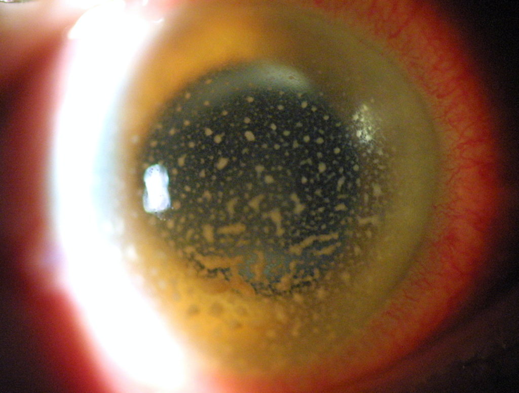
- Cells return, or flare increases, when the dose is reduced
- The patient develops pain, redness, or photophobia
- New keratic precipitates or synechiae appear
When a relapse occurs, return to the previous dose that controlled the eye, and taper more slowly.
Monitoring Pressure and Other Effects
- Measure the pressure at each visit, and at the start and the end of the course
- Watch for cataract with prolonged use
- In patients with frequent relapses, consider systemic therapy to reduce steroid use (see steroid-sparing immunosuppression)
Patient Advice
Explain that patients must use drops as prescribed and must not stop when the eye feels better.
Give a written schedule, and tell the patient to return if symptoms come back.
Check understanding at each visit.
Special Points on Dosing
- Clinicians may combine frequent drops over the first hours with a long-acting periocular injection in severe cases
- Soft steroids, such as loteprednol or fluorometholone, are used for mild disease or for prolonged tapers where pressure rise is a concern
- Ointment at night may improve adherence in some patients
- Suspect a steroid response when pressure rises, and reduce or change the drug
A Sample Case
A 35-year-old man with HLA-B27 disease has 3+ cells and photophobia.
He starts prednisolone acetate every two hours and cyclopentolate, and returns in four days with 1+ cells.
The drops are reduced to six times daily for a week, then four times for a week, then three, two, and once daily over the next three weeks, with review at each step.
At the end, the eye is quiet, and he is advised to return at the first sign of redness.
Recurrent Disease
A patient with two or more attacks a year should have a written plan that allows early treatment, and may need prophylaxis with a systemic drug.
Explain that short courses of early treatment prevent the need for stronger therapy, and keep a record of the dates and the response.


Document what you see
Two smartphone imaging tools built for everyday clinic use — one for the slit lamp, one for the fundus.
From Choroida — the team behind this siteReferences
- Jabs DA, Nussenblatt RB, Rosenbaum JT, et al. Standardization of uveitis nomenclature for reporting clinical data: results of the First International Workshop. Am J Ophthalmol. 2005;140:509-516.
- Agrawal RV, Murthy S, Sangwan V, Biswas J. Current approach in diagnosis and management of anterior uveitis. Indian J Ophthalmol. 2010;58:11-19.
- Foster CS, Vitale AT. Diagnosis and Treatment of Uveitis. 2nd ed. Jaypee; 2013.
- American Academy of Ophthalmology. Basic and Clinical Science Course, Section 9: Uveitis and Ocular Inflammation. AAO; current edition.

Writes and reviews the clinical articles on Educate. Every post is checked against peer-reviewed sources before it goes live.