
Macular coloboma is a congenital, sharply demarcated, excavated defect of the retina and choroid at the macula.
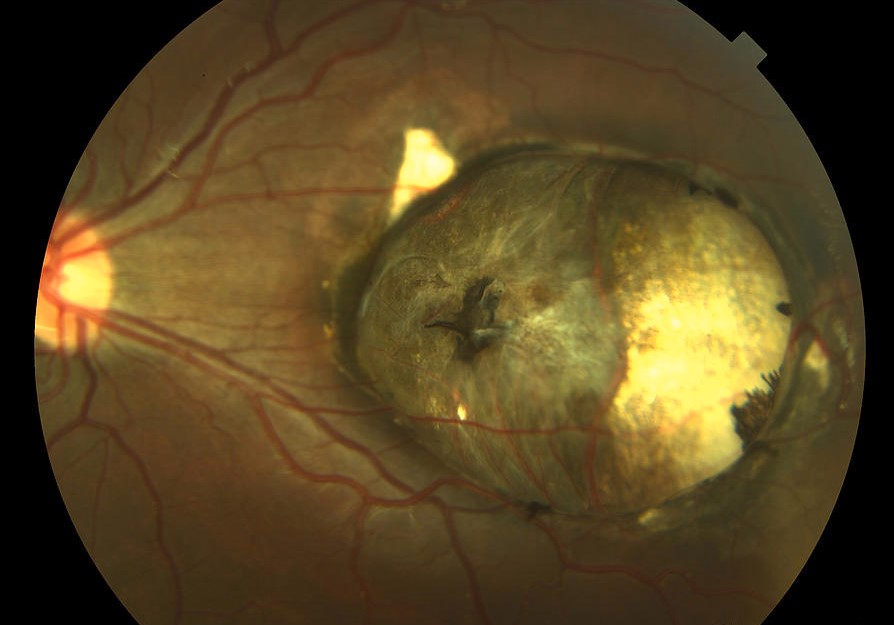
It causes central visual loss from birth, and it is often bilateral.
The lesion looks similar to a scar from toxoplasmosis or to a dystrophy, so the diagnosis depends on history, family examination, and imaging.
What Is Macular Coloboma?
The term is used for a punched-out lesion centered on the macula, and it is not a true coloboma in the embryologic sense because it does not arise from failure of closure of the embryonic fissure.
It is thought to result from a developmental defect, an intrauterine inflammation, or a genetic disorder.
Some cases are familial, with autosomal dominant inheritance in several families, and others are sporadic.
The condition overlaps with the severe form of North Carolina macular dystrophy, which can look the same.
Clinical Features
- A round or oval, well-defined, excavated lesion with a white base of sclera, and a rim of pigment
- Retinal vessels that may cross the base of the lesion
- Reduced central vision from childhood, often 20/200 or worse, and sometimes better than the appearance suggests
- Nystagmus and strabismus when the lesions are bilateral
- Stable appearance over time, with little progression
The lesion is usually 1 to 3 disc diameters in size, and the surrounding retina is normal.
The visual field shows a dense central scotoma.
Fundus Explorer Pro
Photograph the retinal findings described here with the phone already in your pocket — 22 D optics and built-in illumination in one handheld unit.
From Choroida — the team behind this siteImaging
OCT shows the absence or thinning of the retinal layers and choroid at the lesion, with an excavation of the sclera and sometimes a schisis-like separation at the edge.
Fundus autofluorescence shows absent signal in the lesion and a normal or slightly irregular signal at the border.
Fluorescein angiography shows the absence of choroidal filling in the excavation, with normal vessels at the margin.
OCT of the optic disc and the other eye helps to look for other malformations.
Differential Diagnosis
- Congenital toxoplasmosis scar: a pigmented macular scar, often with satellite lesions in the periphery (see ocular toxoplasmosis)
- North Carolina macular dystrophy: an inherited, nonprogressive dystrophy, with a grade that mimics coloboma (see North Carolina macular dystrophy)
- Torpedo maculopathy: a temporal, comet-shaped lesion (see torpedo maculopathy)
- Central areolar choroidal dystrophy and Stargardt disease: progressive macular atrophy (see central areolar choroidal dystrophy)
- True colobomas of the optic disc and the inferior retina: a different location (see coloboma)
Evaluation
Ask about the pregnancy, maternal infections, and family history, and examine the parents and siblings.
Serology for toxoplasma, cytomegalovirus, and other congenital infections is reasonable in an infant with unexplained macular lesions.
Consider genetic testing when there is a family history, because it can distinguish dystrophies from congenital defects.
Management
There is no treatment to restore the lost tissue, so management focuses on function.
Correct refractive error, treat amblyopia where a part of the visual field can be used, and provide low-vision aids and educational support.
Check for retinal detachment and choroidal neovascularization if vision suddenly worsens, although these complications are uncommon.
Counsel the family about the cause, the stable nature of the lesion, and the recurrence risk if the case is familial.
Visual Prospects
Many patients have better vision than the appearance suggests, since some function remains in the tissue around the lesion and eccentric fixation can be used.
Vision in an eye with a large lesion may be limited to counting fingers or hand movements, and in a smaller lesion, it may reach 20/100.
Visual acuity tends to remain stable over decades, and sudden loss should prompt a search for a complication such as a retinal detachment, a macular hole at the edge, or a neovascular membrane.
Familial and Genetic Evaluation
Examine both parents and siblings, because mild forms of the same condition may be found.
Dominant inheritance is the most common pattern when the condition is familial, and the risk to each child of an affected parent is 50 percent.
Genetic testing can identify some forms of macular dystrophy that mimic coloboma, and it can end a diagnostic odyssey for the family.
Associated Findings
Other ocular anomalies, such as microphthalmos, optic disc anomalies, and strabismus, may be present, and a complete examination of both eyes is needed.
In infants with bilateral macular lesions and systemic signs, consider a syndrome and refer to a pediatrician or geneticist.
Counselling
Give the parents an honest and balanced explanation: the lesion is present from birth, it does not grow, and vision will usually be limited but stable.
Early support for visual development, school adaptations, and low-vision devices make a real difference.
Remind them that regular eye examinations are needed to correct refractive error and to monitor for complications.


Document what you see
Two smartphone imaging tools built for everyday clinic use — one for the slit lamp, one for the fundus.
From Choroida — the team behind this siteReferences
- Small KW, Killian J, McLean WC. North Carolina’s dominant progressive foveal dystrophy: how progressive is it? Br J Ophthalmol. 1991;75:401-406.
- Salmon JF. Kanski’s Clinical Ophthalmology: A Systematic Approach. 9th ed. Elsevier; 2020.
- Ryan SJ, Schachat AP, Wilkinson CP, et al, eds. Retina. 6th ed. Elsevier; 2018.
- American Academy of Ophthalmology. Basic and Clinical Science Course, Section 12: Retina and Vitreous. AAO; current edition.

Writes and reviews the clinical articles on Educate. Every post is checked against peer-reviewed sources before it goes live.


