Metaherpetic ulcer is a persistent corneal epithelial defect that follows herpes simplex keratitis after active viral replication has stopped.
Clinicians also call it a trophic or neurotrophic herpetic ulcer.
Because the defect resembles ongoing infection, many eyes receive more antiviral drops.
However, that reflex often delays healing.
What Is a Metaherpetic Ulcer?
The lesion reflects failed healing, and live virus does not drive it.
Earlier infection damages corneal nerves, epithelium and basement membrane.
As a result, new epithelium cannot cover the defect or stay attached.
The ulcer then persists for weeks, although viral testing is usually negative.
Why a Metaherpetic Ulcer Persists
First, herpes damages corneal nerves, which normally send trophic signals to the epithelium.
Confocal microscopy shows reduced subbasal nerve density in these eyes, and this loss tracks with reduced sensation.
Second, poor sensation lowers reflex tearing and blinking.
Finally, basement membrane damage weakens epithelial adhesion.
All-fit Slit-Lamp Adapter
Record and share exactly what you see at the slit lamp. One adapter fits any slit lamp or surgical microscope — and any smartphone.
From Choroida — the team behind this siteThe Role of Drop Toxicity
Prolonged topical antivirals can injure the epithelium and slow cell migration.
Preservatives add further surface toxicity, especially with frequent dosing.
Also ask directly about topical anesthetic use, because anesthetic abuse produces a similar nonhealing defect.
What the Patient Reports
Symptoms are often milder than the slit lamp appearance suggests.
Because sensation is reduced, a large defect may cause little pain.
Patients usually describe blur, redness or mild irritation, plus a history of recurrent herpes keratitis.
However, new severe pain suggests superinfection or another process.
Slit Lamp Signs of Metaherpetic Ulcer
The defect is typically round or oval, with smooth, gray, thickened margins.
It often sits centrally or in the interpalpebral cornea, sometimes at the site of an earlier dendrite.
Fluorescein fills the bed, while the rolled edge may look heaped.
In addition, the ulcer tends to stay static between visits.
Metaherpetic Ulcer vs Active Epithelial Keratitis
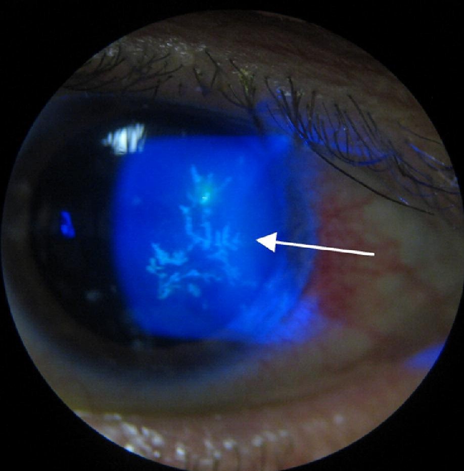
Active herpetic lesions look different.
Dendrites show terminal bulbs, and geographic ulcers have scalloped edges with swollen, virus-laden epithelium at the border.
In contrast, the metaherpetic margin is smooth and lacks terminal bulbs.
Active epithelial disease also tends to improve on antivirals (see herpes simplex keratitis).
Tests That Clarify the Diagnosis
Test corneal sensation in both eyes before instilling anesthetic.
A cotton wisp works at the bedside, while a Cochet-Bonnet esthesiometer gives a graded value.
PCR from the ulcer edge helps when active virus remains possible.
What Must Be Excluded
A stromal infiltrate, purulent discharge or hypopyon suggests bacterial or fungal superinfection.
Therefore, scrape and culture before changing therapy (see why keratitis treatment fails).
Also consider herpes zoster, exposure, lagophthalmos, diabetes and prior corneal surgery, since each can impair healing.
How Treatment of Metaherpetic Ulcer Differs
Active epithelial keratitis needs antiviral therapy or debridement.
By contrast, this ulcer needs a protected surface and time to re-epithelialize.
First, stop unnecessary drops and taper prolonged topical antivirals.
Lubrication and Surface Protection
Preservative-free lubricants and ointment reduce friction and support migrating cells.
A bandage contact lens can also shield the new epithelium from the lids.
However, it carries infection risk in an insensate cornea, so prophylaxis and close review are advisable.
Antiviral Prophylaxis in Metaherpetic Ulcer
Oral antivirals do not close the defect directly.
However, the Herpetic Eye Disease Study showed that oral acyclovir 400 mg twice daily reduced ocular herpes recurrences over 12 months.
Recurrences occurred in 19% of treated patients and 32% of placebo patients.
Thus, many clinicians use prophylaxis during healing, although direct evidence here is limited.
Steroids: Use With Caution
Residual stromal inflammation can slow healing and promote melting.
A low-dose steroid, given under antiviral cover, may help selected eyes.
However, steroids also inhibit wound repair and raise the risk of stromal lysis.
Therefore, reserve them for clear inflammation and review these patients often.
Advanced Therapy for Metaherpetic Ulcer
Autologous serum drops supply growth factors and other epitheliotrophic factors.
Amniotic membrane provides a substrate and has anti-inflammatory effects.
Cenegermin, a recombinant human nerve growth factor, improved healing of moderate and severe neurotrophic keratitis in the REPARO trial.
However, cost limits access, and evidence specific to herpetic ulcers remains limited.
Lid Procedures and Surgery
Tarsorrhaphy or botulinum-induced ptosis protects a cornea that keeps failing.
For impending perforation, tissue adhesive or a patch graft can preserve the globe.
Then, optical keratoplasty should wait because a denervated, previously herpetic cornea heals poorly.
Follow-Up and Counseling for Metaherpetic Ulcer
Review the eye frequently until the epithelium closes, then space visits gradually.
Tell patients that this ulcer usually reflects poor healing, so extra antiviral drops will not speed recovery.
Also warn them that new pain, discharge or sudden vision loss needs urgent review.
Pitfalls to Avoid
- Do not label every persistent defect as active herpes
- Avoid prolonged topical antivirals without a clear indication
- Never skip sensation testing before anesthetic drops


Document what you see
Two smartphone imaging tools built for everyday clinic use — one for the slit lamp, one for the fundus.
From Choroida — the team behind this siteReferences
- Hamrah P, Cruzat A, Dastjerdi MH, et al. Corneal sensation and subbasal nerve alterations in patients with herpes simplex keratitis: an in vivo confocal microscopy study. Ophthalmology. 2010;117:1930-1936.
- Herpetic Eye Disease Study Group. Acyclovir for the prevention of recurrent herpes simplex virus eye disease. N Engl J Med. 1998;339:300-306.
- Bonini S, Lambiase A, Rama P, et al. Phase II randomized, double-masked, vehicle-controlled trial of recombinant human nerve growth factor for neurotrophic keratitis. Ophthalmology. 2018;125:1332-1343.
- Dua HS, Said DG, Messmer EM, et al. Neurotrophic keratopathy. Prog Retin Eye Res. 2018;66:107-131.
Test yourself
A few questions straight from this article.
-
What underlies a metaherpetic ulcer after herpes simplex keratitis?
Live virus does not drive this ulcer: earlier infection damages nerves, epithelium and basement membrane, so new epithelium cannot cover the defect or stay attached. -
How does corneal nerve damage promote a metaherpetic ulcer?
Corneal nerves normally send trophic signals to the epithelium, and poor sensation also lowers reflex tearing and blinking. -
What does confocal microscopy show in eyes with metaherpetic ulcer?
Confocal microscopy shows reduced subbasal nerve density in these eyes, and the loss tracks with reduced corneal sensation. -
What is the typical slit lamp appearance of a metaherpetic ulcer?
The defect is typically round or oval with smooth, gray, thickened margins, often central or interpalpebral, and it tends to stay static between visits. -
Which border marks a geographic herpetic ulcer as active disease rather than a metaherpetic ulcer?
Geographic ulcers have scalloped edges with swollen, virus-laden epithelium at the border, whereas the metaherpetic margin is smooth. -
What is the first treatment step in a metaherpetic ulcer?
This ulcer needs a protected surface and time to re-epithelialize, and more antiviral drops can injure the epithelium and slow cell migration. -
What is the main risk of a bandage contact lens in a metaherpetic ulcer?
The lens shields new epithelium from the lids, but it carries infection risk in an insensate cornea, so close review is advised. -
When should a low-dose steroid be used in a metaherpetic ulcer?
Steroids may help selected eyes under antiviral cover, but they inhibit wound repair and raise the risk of stromal lysis, so they are reserved for clear inflammation with frequent review. -
What is cenegermin, one of the advanced options for a metaherpetic ulcer?
Cenegermin is a recombinant human nerve growth factor that improved healing of moderate and severe neurotrophic keratitis in REPARO, though evidence specific to herpetic ulcers remains limited. -
A presumed metaherpetic ulcer develops a stromal infiltrate and hypopyon. What should come before changing therapy?
A stromal infiltrate, purulent discharge or hypopyon suggests bacterial or fungal superinfection, so the ulcer should be scraped and cultured before therapy changes.

Writes and reviews the clinical articles on Educate. Every post is checked against peer-reviewed sources before it goes live.