Dark adaptation testing measures how quickly the eye regains sensitivity to dim light after exposure to bright light.
Also, slowed recovery is one of the earliest functional signs of age-related macular degeneration.
It is also a key feature of night-blindness disorders and vitamin A deficiency.
The test has long been a laboratory tool, and automated devices have brought it into the clinic.
What Is Dark Adaptation Testing?
When a person walks from daylight into a dark room, vision is poor at first and then improves over many minutes.
The process has two phases: cones recover in the first few minutes, and rods take over and continue to improve for 30 minutes or more.
Rod recovery depends on the regeneration of the visual pigment, rhodopsin, which requires a supply of vitamin A from the retinal pigment epithelium and the choroid (see electroretinography).
Anything that impairs this supply, such as a disease of Bruch membrane or the pigment epithelium, slows the recovery.
Dark Adaptation Testing: How the Test Is Done
- The patient is dilated and exposed to a bright light that bleaches a known proportion of the photopigment
- The examiner places the patient in darkness and asks them to detect a flickering or steady light at a given retinal location
- Then the device dims the light until the patient can no longer see it, and plots the threshold against time
- The curve shows the cone phase, the cone-rod break, and the rod phase
Classical instruments, such as the Goldmann-Weekers adaptometer, measure the whole curve over 30 to 40 minutes.
Modern devices measure the time that it takes for the threshold to fall to a fixed level in the rod phase, called the rod intercept time.
A shorter session can complete the test.
Fundus Explorer Pro
Photograph the retinal findings described here with the phone already in your pocket — 22 D optics and built-in illumination in one handheld unit.
From Choroida — the team behind this siteRod Intercept Time
The rod intercept time is the number of minutes after the bleach until rod sensitivity recovers to a defined criterion.
In healthy older adults it is usually several minutes. In patients with early AMD it is also prolonged, sometimes to 20 minutes or more, which is the maximum time in some tests.
Some protocols regard a delay of more than about 6.5 minutes as abnormal, though the cutoff depends on the device and the age of the patient.
The measure is made in the macula, often 5 degrees from the fovea, where rods are numerous and early AMD changes are present.
Dark Adaptation Testing: Early AMD
Delayed dark adaptation is one of the earliest signs of AMD, and it may precede visible drusen on examination.
Owsley and colleagues showed that delayed rod-mediated adaptation is a functional biomarker for incident early AMD in older adults (see the references).
Studies of diagnostic accuracy found that dark adaptometry could separate patients with early AMD from controls with good sensitivity and specificity (see the references).
The test may identify patients at risk before structural changes are obvious.
Researchers and some clinics use it for this purpose (see age-related macular degeneration).
Other Causes of Delayed Dark Adaptation
- Vitamin A deficiency: reversible with supplementation (see Bitot spots and vitamin A deficiency)
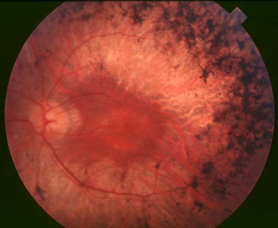
- Retinitis pigmentosa and other rod-cone dystrophies, with a raised threshold (see retinitis pigmentosa)
- Congenital stationary night blindness (see congenital stationary night blindness)
- Oguchi disease and fundus albipunctatus, with very slow recovery (see Oguchi disease)
- Diabetic retinopathy, retinal vascular disease, and other macular disease
- Drugs, liver disease, and malabsorption that lower vitamin A
Because many causes exist, interpret the result with the history and the examination.
Practical Use and Limits
- The test needs cooperation and a quiet room, and takes 20 to 30 minutes
- It is affected by media opacities, pupil size, and fixation
- Normal values vary with age, so use age-related norms
- It does not diagnose AMD alone, and a normal result does not exclude disease
- Cost and availability limit its use
It can help in patients with poor night vision and a normal-looking fundus, and as an adjunct in patients with drusen.
Counselling
Explain that the test checks how the retina recovers after bright light, that it takes some time, and that it gives information that is not available from images.
If the result is abnormal, discuss the steps that can protect the macula, such as not smoking and a healthy diet (see age-related macular degeneration risk factors).
Interpreting a Slow Result in Practice
Ask a patient with a prolonged rod intercept time and a normal-looking macula about vitamin A intake, liver disease, malabsorption, and medications, since a treatable cause may be present.
If there is no such cause and the patient is over 60, early AMD is a leading possibility.
Do an OCT with attention to drusen and reticular pseudodrusen.
Follow-up in six to twelve months can show whether the delay is progressing.
Clinicians can advise the patient about the Amsler grid and risk factors.


Document what you see
Two smartphone imaging tools built for everyday clinic use — one for the slit lamp, one for the fundus.
From Choroida — the team behind this siteReferences
- Owsley C, McGwin G Jr, Clark ME, et al. Delayed rod-mediated dark adaptation is a functional biomarker for incident early age-related macular degeneration. Ophthalmology. 2016;123:344-351.
- Jackson GR, Scott IU, Kim IK, et al. Diagnostic sensitivity and specificity of dark adaptometry for detection of age-related macular degeneration. Invest Ophthalmol Vis Sci. 2014;55:1427-1431.
- Ryan SJ, Schachat AP, Wilkinson CP, et al, eds. Retina. 6th ed. Elsevier; 2018.
- American Academy of Ophthalmology. Basic and Clinical Science Course, Section 12: Retina and Vitreous. AAO; current edition.

Writes and reviews the clinical articles on Educate. Every post is checked against peer-reviewed sources before it goes live.