
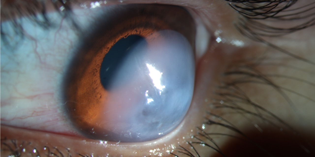
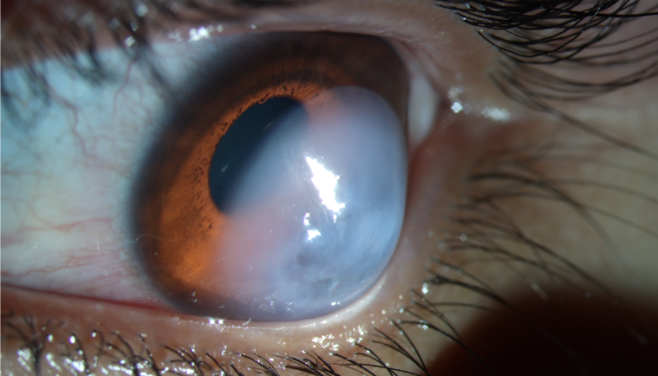
Acute corneal hydrops produces one of the more dramatic sudden changes in corneal appearance a patient can experience: a cornea that looked merely irregular the day before becomes, over hours, densely white and swollen, and understanding the mechanism behind this abrupt transformation, a torn Descemet membrane, explains both the alarming presentation and the generally reassuring natural history that follows.
What Happens Mechanically
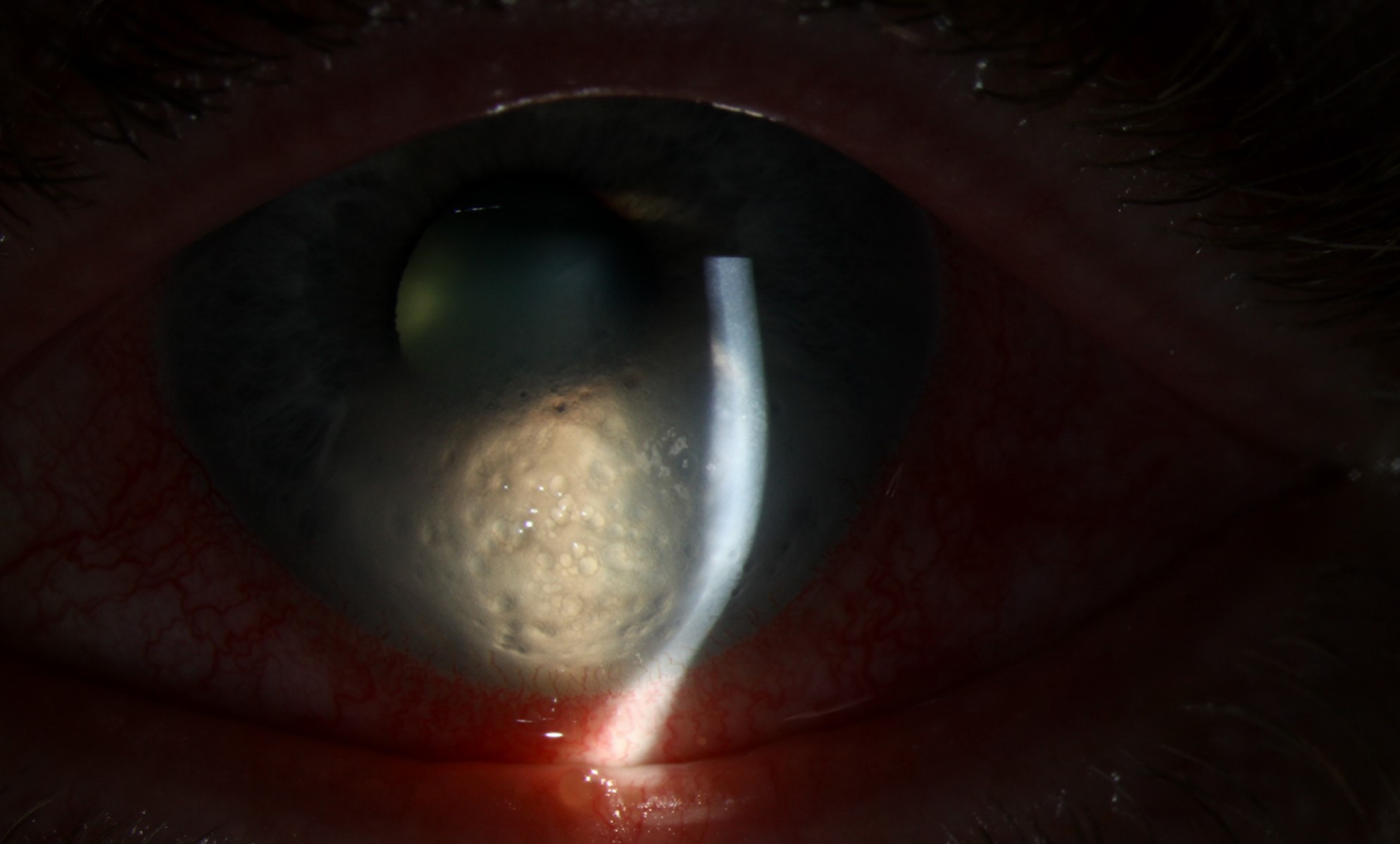
In advanced ectatic corneal disease, the progressive thinning and steepening of the cornea places significant mechanical stress on Descemet membrane, and when this membrane finally ruptures, aqueous humor rushes into the stroma through the break, producing sudden, often dramatic stromal and epithelial edema, since the endothelial pump function normally keeping the stroma relatively dehydrated is abruptly lost in the area of the break.
Underlying Conditions
Acute hydrops occurs almost exclusively in eyes with advanced corneal ectasia, most commonly keratoconus, though it can also occur in pellucid marginal degeneration and other ectatic corneal conditions where the same progressive thinning and mechanical stress on Descemet membrane are present (see pellucid marginal degeneration).
Eye rubbing, a recognized risk factor for keratoconus progression more generally, may also contribute to precipitating an acute hydrops episode in a susceptible, already advanced cornea.
All-fit Slit-Lamp Adapter
Record and share exactly what you see at the slit lamp. One adapter fits any slit lamp or surgical microscope — and any smartphone.
From Choroida — the team behind this siteClinical Presentation
- Sudden onset of significant, often dense, corneal clouding, typically developing over hours to a day or two
- Pain and photophobia, reflecting the acute epithelial and stromal disruption, and reduced vision proportional to the degree of edema
- Marked stromal thickening on examination, sometimes with visible microcystic epithelial edema overlying the area of stromal swelling
- A history of known or suspected keratoconus, or, in some cases, hydrops as the first presentation revealing previously undiagnosed advanced ectatic disease
- The area of Descemet membrane break itself may be visible on careful slit-lamp examination as a discrete, scrolled edge once the acute edema has settled somewhat
Diagnosis
Diagnosis is usually straightforward based on the sudden, dramatic clinical presentation in the setting of known or clinically apparent keratoconus, though anterior segment optical coherence tomography can help confirm the diagnosis by directly visualizing the Descemet membrane break and the extent of stromal fluid, and is useful for monitoring resolution over time.
Management
Conservative Management
The great majority of acute hydrops episodes are managed conservatively, since the endothelium at the margins of the Descemet break gradually migrates to cover the defect and reestablish the pump function, allowing the edema to resolve over a period of weeks to a few months without surgical intervention.
- Hypertonic saline drops and ointment can help reduce epithelial edema and improve comfort during the resolution period
- A bandage contact lens can improve comfort and protect the epithelium during healing
- Topical cycloplegia and, in some regimens, a short course of topical corticosteroid can help with comfort and reduce inflammation, though corticosteroid use is approached cautiously given the already compromised, thinned corneal tissue
- Oral analgesics support pain control during the acute, most symptomatic phase
Intracameral Gas Injection
For larger or more persistent Descemet membrane breaks, injection of air or a longer-acting gas into the anterior chamber can help tamponade the break, pressing the separated edges of Descemet membrane back together and potentially speeding resolution of the hydrops compared with conservative management alone.
What Happens After Resolution
Once the acute hydrops resolves, the affected area of cornea typically develops some degree of permanent stromal scarring at the site of the healed Descemet break, which can, somewhat counterintuitively, sometimes result in modest corneal flattening and a degree of improved contact lens tolerance in that area, even as the scar itself may reduce best corrected visual acuity to some degree.
Long-Term Management
Once the acute episode has resolved and any residual scarring has stabilized, the underlying ectatic disease and its consequences are managed using standard approaches for advanced keratoconus, including consideration of corneal transplantation, generally deep anterior lamellar keratoplasty when the endothelium and Descemet membrane in the non-affected areas remain viable, for eyes with vision-limiting scarring or irregularity (see deep anterior lamellar keratoplasty).
Prognosis
The majority of acute hydrops episodes resolve with conservative management over weeks to a few months, and many patients regain reasonable, sometimes surprisingly good, vision once the acute edema clears, even though some residual scarring is typical.
Corneal crosslinking is not applicable during an acute hydrops episode itself, since the priority is managing the acute edema, but the episode itself often marks a point at which corneal transplantation becomes a more actively considered option, given that hydrops generally reflects advanced, severe underlying ectatic disease.


Document what you see
Two smartphone imaging tools built for everyday clinic use — one for the slit lamp, one for the fundus.
From Choroida — the team behind this siteReferences
- Fan Gaskin JC, Patel DV, McGhee CN. Acute corneal hydrops in keratoconus – new perspectives. Am J Ophthalmol. 2014;157:921-928.
- Grewal S, Laibson PR, Cohen EJ, Rapuano CJ. Acute hydrops in the corneal ectasias: associated factors and outcomes. Trans Am Ophthalmol Soc. 1999;97:187-198.
- Miller DD, Hasan SA, Simmons NL, Stewart MW. Recurrent corneal erosion: a comprehensive review. Clin Ophthalmol. 2019;13:325-336.
- Panda A, Aggarwal A, Madhavi P, et al. Management of acute corneal hydrops secondary to keratoconus with intracameral injection of sulfur hexafluoride (SF6). Cornea. 2007;26:1067-1069.
