
Electroretinography (ERG) records the electrical response of the retina to a light stimulus, and it is the objective test of retinal function that fills the gap when the fundus looks normal and the patient cannot see.
It separates rod from cone dysfunction, outer from inner retinal disease, and generalized from macular disease.
Understanding the standard responses, and what each waveform component represents, allows the ophthalmologist to request the right test and to read the report critically.
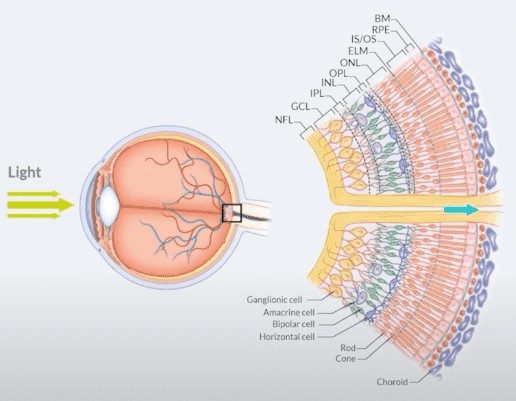
What Electroretinography Measures
The full-field ERG is generated by the whole retina in response to a flash.
- A-wave: the initial negative deflection, generated by photoreceptors
- B-wave: the positive deflection that follows, generated by ON-bipolar cells with contribution from Müller cells
- Oscillatory potentials: small wavelets on the rising b-wave, related to inner retinal activity including amacrine cells
- Photopic negative response: a slow negative wave after the b-wave in the light-adapted ERG, related to retinal ganglion cell activity
The ERG does not measure ganglion cell function directly except through the photopic negative response and the pattern ERG, and it does not test the optic nerve or visual cortex.
The Standard Full-Field ERG
The International Society for Clinical Electrophysiology of Vision (ISCEV) defines the standard protocol.
Patients are dark-adapted for at least 20 minutes and then tested in the dark and after light adaptation.
- Dark-adapted 0.01 ERG: a dim flash producing a b-wave that reflects rod-driven activity
- Dark-adapted 3.0 ERG (standard combined response): a bright flash producing a mixed rod and cone response with a clear a-wave and b-wave
- Dark-adapted 10 ERG: a brighter flash that emphasizes the a-wave and photoreceptor function
- Light-adapted 3.0 ERG: a cone-driven response
- Light-adapted 30 Hz flicker ERG: a cone-pathway response, sensitive to cone dysfunction
Recording uses a corneal contact electrode or a conjunctival fiber electrode, and skin electrodes can be used in young children with lower amplitudes.
Pupils are dilated, and the results are compared with normative data from the laboratory.
Fundus Explorer Pro
Photograph the retinal findings described here with the phone already in your pocket — 22 D optics and built-in illumination in one handheld unit.
From Choroida — the team behind this siteInterpreting the Results
Rod-Cone Pattern
Rod responses are reduced more than cone responses in retinitis pigmentosa and related disorders.
The DA 0.01 response is affected early, and later the cone responses are reduced (see retinitis pigmentosa).
Cone-Rod and Cone Pattern
Cone-rod dystrophies show reduced cone responses first (see cone-rod dystrophy).
Achromatopsia shows absent or markedly reduced photopic and flicker responses with normal rod responses.
Negative ERG
A negative ERG is one in which the b-wave is smaller than the a-wave in the standard combined response.
It indicates dysfunction of the post-photoreceptor circuitry, and causes include:
- Congenital stationary night blindness, complete and incomplete types (see congenital stationary night blindness)
- X-linked retinoschisis (see X-linked retinoschisis)
- Central retinal artery occlusion and severe retinal ischemia
- Melanoma-associated retinopathy and paraneoplastic disease
- Some toxicities, such as quinine
- Batten disease and other neuronal storage disorders
Normal Full-Field ERG With Vision Loss
A normal full-field ERG does not exclude macular disease, because the macula contributes only a small portion of the total response.
In such cases, multifocal ERG or pattern ERG may show a focal or macular abnormality.
Multifocal and Pattern ERG
Multifocal ERG
The multifocal ERG (mfERG) maps cone-driven responses across the central retina, typically within 30 degrees.
It shows focal loss of function in macular disease, toxic maculopathy, and occult macular dystrophy.
It can reveal early hydroxychloroquine damage when other tests are equivocal, and it is one of the recommended objective tests in screening guidelines.
Pattern ERG
The pattern ERG uses a reversing checkerboard and reflects macular and retinal ganglion cell function.
It helps separate macular disease from optic neuropathy when acuity is reduced.
Both tests require good fixation and refractive correction, and both have standards published by ISCEV.
When to Request an ERG
- Suspected inherited retinal disease, including night blindness, ring scotoma, or family history
- Unexplained visual loss with a normal or minimally abnormal fundus
- Suspected cone or macular disease in a child
- Suspected autoimmune or paraneoplastic retinopathy
- Toxic retinopathy, such as vigabatrin, hydroxychloroquine, or deferoxamine
- Evaluation of retinal function behind opaque media
- Unexplained nystagmus or poor vision in infancy, where Leber congenital amaurosis, achromatopsia, and congenital stationary night blindness are possible
- Monitoring of progression in some genetic diseases and in clinical trials
Worked Examples
- A child with poor vision and nystagmus, normal fundus: absent photopic and flicker responses with normal rod responses points to achromatopsia
- An adult with night blindness and a normal fundus: absent or very small dark-adapted responses with a negative waveform suggests congenital stationary night blindness
- A patient with progressive central vision loss, normal full-field ERG, and reduced multifocal responses centrally: macular dystrophy or toxic maculopathy is likely
- A patient with sudden bilateral photopsia and field loss with a normal fundus: reduced responses in a full-field ERG support an autoimmune or paraneoplastic retinopathy
Each pattern narrows the differential and directs genetic or systemic testing.
Limitations and Practical Points
ERG amplitude varies with age, refractive error, pupil size, electrode type, and laboratory.
Interpretation needs laboratory normal ranges and an experienced reader.
Young children often require sedation or general anesthesia for reliable recordings, so coordination with the anesthesia team is needed.
Media opacity reduces the response, but ERG can still be recorded through cataract and vitreous hemorrhage, unlike many imaging tests.
An ERG result never stands alone.
It is combined with history, examination, imaging, and often genetic testing to reach the diagnosis.


Document what you see
Two smartphone imaging tools built for everyday clinic use — one for the slit lamp, one for the fundus.
From Choroida — the team behind this siteReferences
- Robson AG, Frishman LJ, Grigg J, et al. ISCEV Standard for full-field clinical electroretinography (2022 update). Doc Ophthalmol. 2022;144:165-177.
- McCulloch DL, Marmor MF, Brigell MG, et al. ISCEV Standard for full-field clinical electroretinography (2015 update). Doc Ophthalmol. 2015;130:1-12.
- Hoffmann MB, Bach M, Kondo M, et al. ISCEV standard for clinical multifocal electroretinography (mfERG) (2021 update). Doc Ophthalmol. 2021;142:5-16.
- Bach M, Brigell MG, Hawlina M, et al. ISCEV standard for clinical pattern electroretinography (PERG): 2012 update. Doc Ophthalmol. 2013;126:1-7.
- Marmor MF, Kellner U, Lai TY, Melles RB, Mieler WF; American Academy of Ophthalmology. Recommendations on screening for chloroquine and hydroxychloroquine retinopathy (2016 revision). Ophthalmology. 2016;123:1386-1394.
Test yourself
A few questions straight from this article.
-
The full-field electroretinogram records the electrical response of which part of the visual system?
The full-field ERG is generated by the whole retina in response to a flash. It does not test the optic nerve or the visual cortex, and it assesses ganglion cells only through the photopic negative response and the pattern ERG. -
Which cells generate the a-wave of the full-field electroretinogram?
The a-wave is the initial negative deflection and is generated by the photoreceptors. The positive b-wave that follows comes from ON-bipolar cells. -
Which cells generate the b-wave of the full-field electroretinogram?
The b-wave is the positive deflection following the a-wave, generated by ON-bipolar cells with a contribution from Müller cells. The small wavelets on its rising limb are the oscillatory potentials. -
How long must a patient be dark-adapted before the ISCEV standard full-field ERG?
The ISCEV protocol requires dark adaptation for at least 20 minutes, after which the patient is tested in the dark and then again after light adaptation, with pupils dilated. -
What does the light-adapted 30 Hz flicker ERG assess?
The light-adapted 30 Hz flicker ERG is a cone-pathway response and is sensitive to cone dysfunction. Rod-driven activity is reflected instead in the dark-adapted 0.01 response. -
What defines a negative ERG?
A negative ERG is one in which the b-wave is smaller than the a-wave in the standard combined response, indicating dysfunction of the post-photoreceptor circuitry. Causes include congenital stationary night blindness and X-linked retinoschisis. -
Which ERG pattern points to achromatopsia?
Achromatopsia shows absent or markedly reduced photopic and flicker responses with normal rod responses, a useful pattern in a child with poor vision, nystagmus and a normal fundus. -
A patient has progressive central vision loss but a normal full-field ERG. Which test is likely to show the abnormality?
A normal full-field ERG does not exclude macular disease, because the macula contributes only a small portion of the total response. Multifocal ERG maps cone-driven responses across the central retina, typically within 30 degrees. -
What does the pattern ERG help distinguish when visual acuity is reduced?
The pattern ERG uses a reversing checkerboard and reflects macular and retinal ganglion cell function, which helps separate macular disease from optic neuropathy. It requires good fixation and refractive correction. -
Which practical advantage does the ERG have over many imaging tests?
Media opacity reduces the response, but an ERG can still be recorded through cataract and vitreous haemorrhage, unlike many imaging tests. Amplitude still varies with age, refraction, pupil size, electrode type and laboratory.

Writes and reviews the clinical articles on Educate. Every post is checked against peer-reviewed sources before it goes live.


