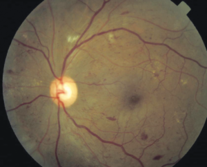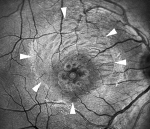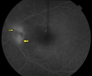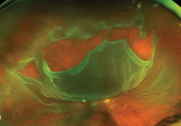
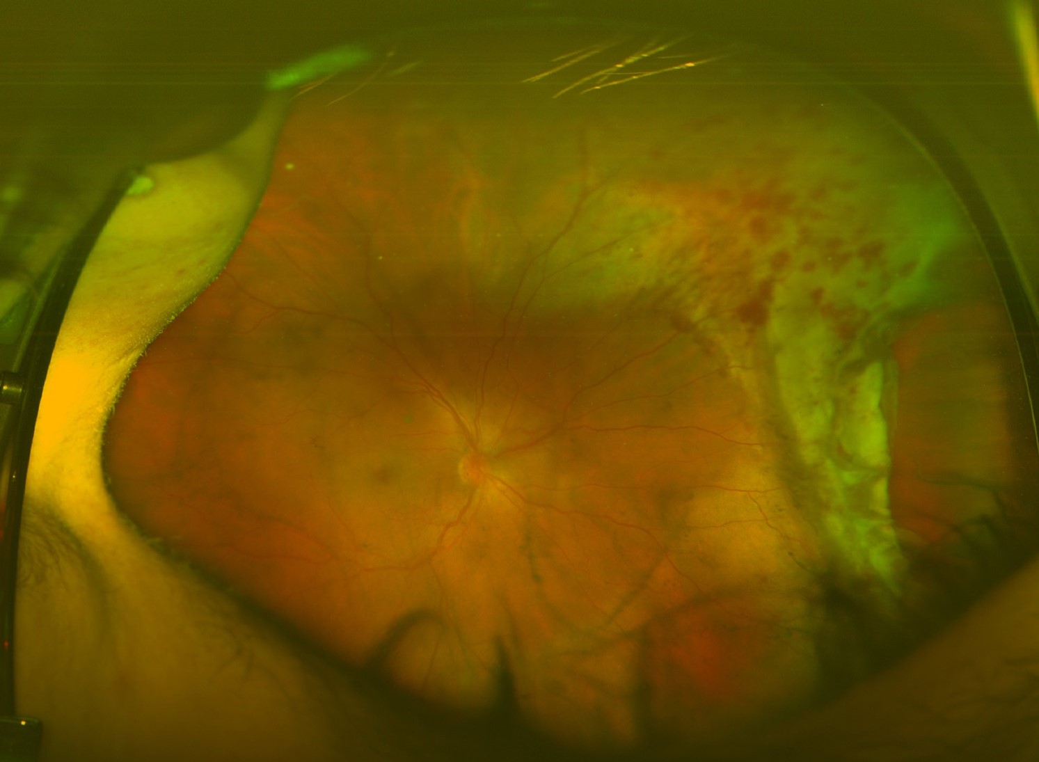
Traumatic retinal detachment is a retinal detachment that follows an injury to the eye.
It differs from the age-related form in its causes, its timing, and its tendency to cause scarring.
It affects young people, mostly men, and it may appear weeks or months after the blow.
Careful follow-up after any significant eye injury is the main defense.
How Trauma Causes Traumatic Retinal Detachment
Blunt trauma compresses the globe in the anteroposterior direction and expands it in the equatorial plane, which stretches the vitreous base.
The traction can also pull the retina away from the ora serrata, producing a dialysis, or tear the retina at other sites, including giant tears.
Penetrating injuries can cause detachment by direct retinal damage, by hemorrhage and scarring, and by contracting vitreous membranes (see intraocular foreign body).
Contusion can also cause macular holes and choroidal rupture, which may lead to detachment in some eyes (see traumatic macular hole and choroidal rupture).
Common Patterns
- Retinal dialysis: a circumferential tear at the ora serrata, typically in the inferotemporal quadrant (see retinal dialysis)
- Giant retinal tear: a tear extending over 90 degrees (see giant retinal tear)
- Vitreous base avulsion: a tear of the vitreous base, often with a bucket-handle appearance (see vitreous base avulsion)
- Macular hole detachment
- Tractional detachment after penetrating injury
Dialysis detachments often progress slowly and may be asymptomatic for a long time, with a demarcation line.
Fundus Explorer Pro
Photograph the retinal findings described here with the phone already in your pocket — 22 D optics and built-in illumination in one handheld unit.
From Choroida — the team behind this siteTiming of Presentation
Clinicians may find a traumatic detachment at the first examination, when the view of the retina is clear.
In many cases, the vitreous hemorrhage or lens opacity hides the retina, and the detachment declares itself later.
Dialyses can take months or years to cause symptoms, and the diagnosis may come long after the injury.
Because of this, patients with significant blunt injuries need dilated examination with scleral depression at the time of injury and at follow-up intervals.
Evaluation for Traumatic Retinal Detachment
- Take a history of the injury, and examine for other signs of trauma
- Dilate the pupil, if there is no open globe, and perform a careful examination of the periphery with indentation
- Use B-scan ultrasonography when the view is hidden (see B-scan ultrasonography)
- Obtain OCT of the macula for a hole or other injury
- Examine the other eye
Look for demarcation lines, pigment, and signs of chronicity, which suggest a slowly progressive detachment.
Treatment for Traumatic Retinal Detachment
Surgery is the standard of treatment for symptomatic or progressive detachment.
- Scleral buckle: often chosen for dialysis in a young patient with an attached vitreous, with cryotherapy or laser (see scleral buckle surgery)
- Pars plana vitrectomy: used for giant tears, tractional components, vitreous hemorrhage, and complex cases (see pars plana vitrectomy)
- Pneumatic retinopexy: occasionally for selected breaks
- Silicone oil or gas tamponade: chosen according to the case
- Laser demarcation: for small, stable dialyses without symptoms
Management of proliferative vitreoretinopathy, which is also more common in trauma, may need membrane peeling and long tamponade (see proliferative vitreoretinopathy).
Prognosis of Traumatic Retinal Detachment
Outcomes depend on the extent of the injury, the damage to the macula and the optic nerve, and the presence of proliferative vitreoretinopathy.
Young patients with a simple dialysis do well, but those with a penetrating injury and multiple complications may have poor vision.
Prompt repair improves the outlook.
Prevention and Counselling
Advise protective eyewear for sports and work, since trauma is largely preventable.
Teach patients who have had a significant blow to report new floaters, flashes, or visual field changes immediately, even months later.
In addition, arrange follow-up examinations, and examine the fellow eye.
Examination Under Anesthesia and Timing
In children and in patients with severe trauma who cannot cooperate, an examination under anesthesia permits a thorough view of the periphery and may be done at the time of other repairs.
Surgery timing depends on the type of injury: surgeons close an open globe injury first.
Surgeons repair a retinal detachment within days or after the early inflammation has settled, according to the case.
In eyes with a clear view and a fresh detachment, early repair gives the best chance, and delay increases the risk of proliferative vitreoretinopathy.
Special Features of Dialysis
Retinal dialysis after trauma tends to occur in the inferotemporal quadrant because the superonasal quadrant is protected by the nose and the brow.
The detachment is often shallow and slow.
Some eyes with a long-standing dialysis and a demarcation line can be observed or treated with laser.
The vitreous is usually still attached, so a scleral buckle is a good option, with minimal risk of cataract in a young patient.
Risks of Missing the Diagnosis
Patients with trauma and visual symptoms months or years later may be misdiagnosed or lost to follow-up, so always ask for a history of trauma in a young patient with a retinal detachment.
In the fellow eye, a careful examination is also needed because some conditions that predispose to trauma-related detachment are bilateral.


Document what you see
Two smartphone imaging tools built for everyday clinic use — one for the slit lamp, one for the fundus.
From Choroida — the team behind this siteReferences
- Ryan SJ, Schachat AP, Wilkinson CP, et al, eds. Retina. 6th ed. Elsevier; 2018.
- Wilkinson CP, Rice TA. Michels Retinal Detachment. 2nd ed. Mosby; 1997.
- Yanoff M, Duker JS, eds. Ophthalmology. 5th ed. Elsevier; 2019.
- American Academy of Ophthalmology. Basic and Clinical Science Course, Section 12: Retina and Vitreous. AAO; current edition.

Writes and reviews the clinical articles on Educate. Every post is checked against peer-reviewed sources before it goes live.