
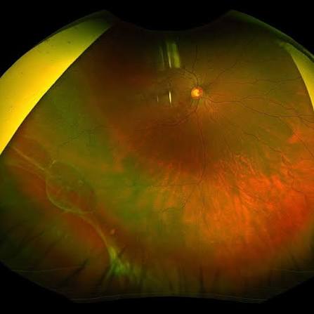
Retinal dialysis is a full-thickness disinsertion of the retina at the ora serrata, a distinct type of peripheral break from the horseshoe tears and atrophic holes covered elsewhere on this site.
It causes a slowly progressive rhegmatogenous detachment that can go unnoticed for weeks or months, which is very different from the acute, symptomatic presentation typical of a horseshoe tear.
That slow tempo, more than any other feature, is what shapes how retinal dialysis actually presents in clinic.
Retinal dialysis accounts for a relatively small proportion of all rhegmatogenous detachments overall, but it makes up a disproportionate share of detachments seen in children and young adults, particularly after trauma.
What Makes Dialysis Different
A dialysis is a circumferential separation of the retina from its normal attachment at the ora serrata, occurring at the vitreous base rather than at a point of focal vitreoretinal traction the way a horseshoe tear does.
Because the break runs along an arc rather than being a single point defect, subretinal fluid tracks in slowly and the resulting detachment tends to advance gradually rather than presenting as a sudden curtain.
The classic location is inferotemporal, and the classic patient is a young, otherwise healthy myope, though the trauma-associated form has its own separate, somewhat different demographic described below.
Etiology
Traumatic dialysis follows blunt ocular trauma, where the sudden globe deformation stretches and tears the vitreous base attachment, and it can occur even without any other visible sign of significant ocular injury.
Spontaneous, non-traumatic dialysis occurs more often in young myopic patients without a clear inciting event, and is thought to relate to an area of abnormally firm vitreoretinal adhesion at the vitreous base that gives way under ordinary eye movement and vitreous traction.
Some cases are associated with specific predisposing conditions affecting the peripheral retina and vitreous base, which is part of why a careful peripheral exam of both eyes matters even when only one eye is symptomatic.
Fundus Explorer Pro
Photograph the retinal findings described here with the phone already in your pocket — 22 D optics and built-in illumination in one handheld unit.
From Choroida — the team behind this siteClinical Presentation
- Painless, gradually enlarging visual field defect, often noticed only once it has advanced significantly toward the macula
- Minimal or absent flashes and floaters compared to a horseshoe tear, since the mechanism does not involve the same acute, focal vitreous traction
- A history of blunt trauma in the traumatic form, sometimes weeks to months before the detachment becomes symptomatic
- Late presentation is common, and it is not unusual for a dialysis-related detachment to be diagnosed only once it threatens or involves the macula
Examination Findings
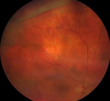
Careful scleral depression around the entire peripheral retina, particularly the inferotemporal quadrant, is essential to identify the characteristic crescent-shaped area of bare, disinserted retinal edge at the ora serrata.
The detached retina is often thin and relatively immobile compared to the more acutely mobile detachment associated with a fresh horseshoe tear, reflecting its slower, more chronic course.
Demarcation lines, pigmented borders that form as a detachment stabilizes over time, are frequently visible in a dialysis-related detachment and are a useful clue to its chronicity even before the diagnosis is otherwise confirmed.
Differential Diagnosis
- Horseshoe tear — a focal, point defect from acute vitreoretinal traction, typically causing a rapidly symptomatic detachment rather than the slow course typical of a dialysis
- Atrophic round hole — usually smaller, often within an area of lattice degeneration, and generally not associated with the same circumferential ora serrata separation
- Giant retinal tear — a much larger circumferential break, but located posterior to the vitreous base rather than at the ora serrata itself, and typically far more acutely symptomatic
- Retinoschisis — a splitting of the retinal layers rather than a full-thickness break, distinguished on careful examination and, when needed, OCT through the peripheral lesion
Management
Scleral buckling is the traditional and still commonly used approach, since the break sits anteriorly and is well suited to an external, segmental or encircling buckle placed under the area of dialysis.
Pars plana vitrectomy is used selectively, particularly when there is associated proliferative vitreoretinopathy, extensive detachment, or other posterior pathology that a buckle alone would not adequately address.
Laser or cryotherapy is applied to the margins of the dialysis to create a chorioretinal adhesion once the retina is repositioned, following the same barrier principle used for any other retinal break.
Because presentation is often delayed, macular involvement at the time of diagnosis is more common than with an acutely symptomatic tear, and this has a direct bearing on the visual prognosis that families need to understand before surgery.
The fellow eye deserves the same careful peripheral examination given at diagnosis, since bilateral dialysis, though uncommon, has been reported, and a young myopic patient with one confirmed dialysis carries some elevated risk in the other eye as well.


Document what you see
Two smartphone imaging tools built for everyday clinic use — one for the slit lamp, one for the fundus.
From Choroida — the team behind this siteReferences
- Wilkinson CP, Rice TA. Michels’ Retinal Detachment.
- American Academy of Ophthalmology. Basic and Clinical Science Course, Section 12: Retina and Vitreous.
- Ross WH. Traumatic retinal dialysis. Archives of Ophthalmology.
- Zion VM, Burton TC. Retinal dialysis: cases with and without antecedent trauma. Archives of Ophthalmology.
Test yourself
A few questions straight from this article.
-
Retinal dialysis is best defined as which of the following?
A dialysis is a full-thickness, circumferential disinsertion of the retina from its attachment at the ora serrata, occurring at the vitreous base rather than at a point of focal traction. -
Which quadrant is the classic site of a retinal dialysis?
The classic location is inferotemporal, which is why careful scleral depression of that quadrant in particular is essential when a dialysis is suspected. -
In which patients does retinal dialysis account for a disproportionate share of retinal detachments?
Dialysis causes only a small proportion of rhegmatogenous detachments overall, but a disproportionate share of those seen in children and young adults, especially after trauma. -
How does blunt ocular trauma produce a retinal dialysis?
Blunt trauma deforms the globe suddenly, stretching and tearing the vitreous base attachment; this can happen without any other visible sign of significant ocular injury. -
How do the symptoms of retinal dialysis differ from those of a horseshoe tear?
Because the mechanism does not involve the same acute focal vitreous traction, flashes and floaters are minimal or absent; patients instead notice a painless, slowly enlarging field defect. -
Which examination technique is essential to identify a retinal dialysis?
Careful scleral depression around the whole periphery, especially inferotemporally, reveals the characteristic crescent of bare, disinserted retinal edge at the ora serrata. -
What do pigmented demarcation lines indicate in a dialysis-related detachment?
Demarcation lines are pigmented borders that form as a detachment stabilises, so their presence is a useful clue to a chronic, slowly progressive course. -
Which feature separates a giant retinal tear from a retinal dialysis?
A giant retinal tear is a much larger circumferential break, but sits posterior to the vitreous base rather than at the ora serrata, and is typically far more acutely symptomatic. -
Why is scleral buckling well suited to repairing a retinal dialysis?
The anterior position of the break suits a segmental or encircling buckle placed under the dialysis; laser or cryotherapy to its margins then creates the chorioretinal adhesion. -
Why is the visual prognosis in retinal dialysis often guarded at diagnosis?
Because the detachment advances slowly and is often noticed late, macular involvement at diagnosis is commoner than with an acutely symptomatic tear, which directly affects visual prognosis.

Writes and reviews the clinical articles on Educate. Every post is checked against peer-reviewed sources before it goes live.


