Venous beading is an irregular, segmental change in the caliber of retinal veins, in which dilated and narrowed segments alternate along the vessel like a string of beads.
In diabetic retinopathy it is also a marker of significant retinal ischemia.
It is also among the most reliable signs that the disease is about to progress.
It is easy to overlook if the examiner looks only at the hemorrhages.
What Is Venous Beading?
The retinal veins normally have a smooth, uniform caliber that changes gradually along their course.
In venous beading, the vein shows focal dilation separated by short segments of narrowing, giving a sausage-like or rosary appearance.
The change reflects damage to the wall and endothelium of the vein in a region of poor perfusion.
It often occurs next to areas of capillary closure.
It may also be accompanied by venous loops, reduplication, and sheathing.
Beading usually appears in the arcades and midperiphery, and it can also affect a single vein or several.
Clinical Features of Venous Beading
- A vein with alternating segments of dilation and narrowing, often with a darker color
- Associated venous loops or reduplication
- Adjacent cotton wool spots, intraretinal hemorrhages, and IRMA (see intraretinal microvascular abnormalities)
- Peripheral capillary nonperfusion on angiography
- A patient with long-standing diabetes who may have few or no symptoms
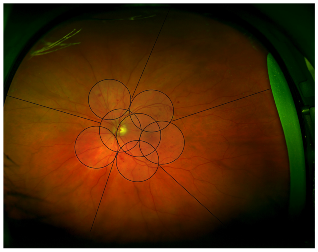
Examination with a red-free filter and with a widefield image makes the finding easier to see (see ultra-widefield retinal imaging).
Fundus Explorer Pro
Photograph the retinal findings described here with the phone already in your pocket — 22 D optics and built-in illumination in one handheld unit.
From Choroida — the team behind this siteWhy It Matters in the Classification
Venous beading in two or more quadrants is one of the three criteria for severe nonproliferative diabetic retinopathy.
Also, the ETDRS found that venous beading was a strong predictor of progression to proliferative retinopathy, and that the risk increased with the extent of the sign.
Also, the 4-2-1 rule uses venous beading as the “2” (see diabetic retinopathy classification).
An eye with beading in two quadrants is therefore at a high risk of developing neovascularization within a year.
The follow-up should reflect that.
Differential Diagnosis of Venous Beading
- Retinal vein occlusion: engorgement, tortuosity, and hemorrhage in a wedge or in the whole fundus (see central retinal vein occlusion)
- Hyperviscosity syndromes: dilated, segmented veins in a patient with a blood disorder (see Waldenström macroglobulinemia)
- Ocular ischemic syndrome: dilated veins, midperipheral hemorrhages, and a history of carotid disease (see ocular ischemic syndrome)
- Retinal vasculitis: sheathing and leakage of vessels, often with inflammation (see retinal vasculitis)
- Sickle cell and other proliferative retinopathies (see sickle cell retinopathy)
A history of diabetes, with other features of diabetic retinopathy in the same eye, makes the diagnosis clear.
Evaluation and Management
Perform a dilated examination of all quadrants and obtain fundus photographs.
In addition, fluorescein angiography can show the extent of nonperfusion and exclude early neovascularization (see fluorescein angiography patterns).
Also, review the patient’s blood glucose control, blood pressure, lipids, and kidney function with the primary physician.
The retinal treatment depends on the whole picture: eyes with severe nonproliferative disease may also be monitored at short intervals or treated with panretinal photocoagulation or anti-VEGF drugs (see panretinal photocoagulation).
In addition, patients should be told that the change in the veins reflects reduced blood supply to the retina, that it can progress, and that regular visits and good systemic control reduce the chance of vision loss.
Beading in Other Conditions
Venous beading is not exclusive to diabetes.
It can appear in retinal vein occlusion, retinal vasculitis, and hyperviscosity states, so the clinical context matters.
In sickle cell and other proliferative retinopathies, sea fan neovascularization at the periphery is associated with other vascular changes.
An isolated bead in a single vein, in an eye without other diabetic changes, is also unlikely to carry the same weight as extensive beading.
Patient Counselling
Tell the patient that the veins in the retina show a pattern linked to reduced blood supply, that this is a warning and not a loss of vision, and that treatment can be started if the disease progresses.
Review the practical steps: keep blood sugar, blood pressure, and cholesterol in range, take every follow-up appointment, and report new floaters or a sudden fall in vision.
Because this finding can move quickly, a missed appointment matters, and a reminder system or a telephone follow-up may help patients who have difficulty attending.
Pay attention to the fellow eye, because beading is usually bilateral and sometimes asymmetric.
A careful record of the quadrants affected provides a baseline that makes later change much easier to recognize.
For the latest studies, see the PubMed results for venous beading.


Document what you see
Two smartphone imaging tools built for everyday clinic use — one for the slit lamp, one for the fundus.
From Choroida — the team behind this siteReferences
- Early Treatment Diabetic Retinopathy Study Research Group. Fundus photographic risk factors for progression of diabetic retinopathy. ETDRS report number 12. Ophthalmology. 1991;98:823-833.
- Wilkinson CP, Ferris FL III, Klein RE, et al. Proposed international clinical diabetic retinopathy and diabetic macular edema disease severity scales. Ophthalmology. 2003;110:1677-1682.
- Ryan SJ, Schachat AP, Wilkinson CP, et al, eds. Retina. 6th ed. Elsevier; 2018.
- American Academy of Ophthalmology. Basic and Clinical Science Course, Section 12: Retina and Vitreous. AAO; current edition.

Writes and reviews the clinical articles on Educate. Every post is checked against peer-reviewed sources before it goes live.