
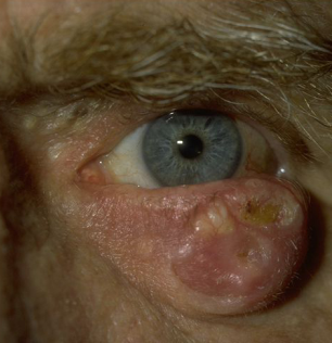
Eyelid squamous cell carcinoma (SCC) is the second most common malignant tumor of the eyelid after basal cell carcinoma, and it behaves more aggressively.
It arises in sun-damaged skin in older patients, and in people who are immunosuppressed or who have inherited sensitivity to ultraviolet light.
Early biopsy and complete excision are the keys to cure.
What Is Eyelid Squamous Cell Carcinoma?
The tumor is a malignant proliferation of keratinocytes, and it often arises from a precursor such as actinic keratosis (see actinic keratosis).
Risk factors include fair skin, chronic sun exposure, advanced age, a history of skin cancer, immunosuppression after organ transplantation, HIV infection, and xeroderma pigmentosum.
The lower eyelid and the lid margin are the most common sites, followed by the medial canthus and the upper lid.
In contrast with basal cell carcinoma, SCC has a recognized capacity for perineural invasion and for spread to regional lymph nodes.
Clinical Features
- A firm, hyperkeratotic nodule or plaque, sometimes ulcerated and crusted
- An irregular, scaly, erythematous patch that fails to heal
- Loss of eyelashes at the lesion, which is a warning sign (see madarosis)
- A fixed, indurated mass with irregular margins
- Pain, numbness, or a nerve palsy when there is perineural spread
The lesion may look like a chalazion, a cutaneous horn, or a benign keratosis, and a lesion that does not follow the expected course should be biopsied.
All-fit Slit-Lamp Adapter
Record and share exactly what you see at the slit lamp. One adapter fits any slit lamp or surgical microscope — and any smartphone.
From Choroida — the team behind this siteDifferential Diagnosis
- Basal cell carcinoma: pearly nodule with telangiectasia and rolled edges (see basal cell carcinoma of the eyelid)
- Sebaceous carcinoma: a masquerade of chalazion or blepharitis (see sebaceous carcinoma)
- Keratoacanthoma: a rapidly growing, crater-shaped lesion (see keratoacanthoma)
- Actinic keratosis and seborrheic keratosis: benign or precursor lesions
- Chronic blepharitis and inflammatory lesions: with a different course
Diagnosis
Biopsy confirms the diagnosis, and it should include the deepest part of the lesion.
Examine the regional lymph nodes in the preauricular and submandibular regions.
When perineural invasion is suspected, MRI of the orbit and skull base with contrast helps to find involvement of the trigeminal or facial nerve.
The pathology report should describe the differentiation, the depth, and the presence of perineural invasion, and these factors affect the prognosis.
Treatment
Surgical excision with controlled margins is the standard of care.
Mohs micrographic surgery or frozen-section margin control allows tissue preservation, which matters on the eyelid, where a small error can affect the function (see Mohs surgery and eyelid reconstruction).
Reconstruction restores the eyelid margin and function, and it is carried out after the margins are clear.
Radiotherapy is an option for patients who cannot have surgery and as an adjunct in cases with perineural invasion or positive margins.
Lymph node disease requires surgery and sometimes radiotherapy.
Recurrence and new skin cancers occur, so lifelong skin examination is advised.
Patients should be told about sun protection, hats, and regular dermatologic checks.
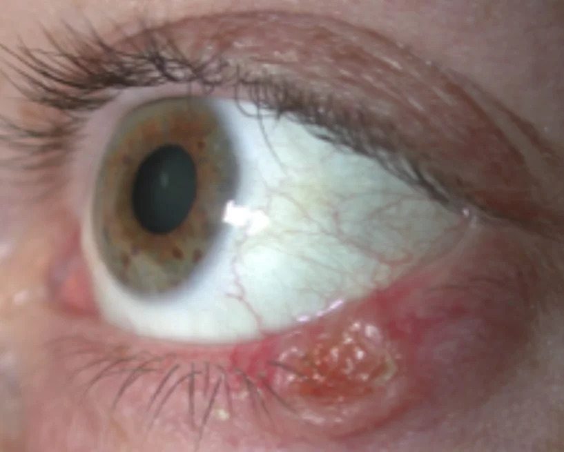
Staging and Prognosis
The prognosis depends on the size of the tumor, the depth of invasion, the degree of differentiation, and the presence of perineural invasion or nodal disease.
Tumors larger than about two centimeters, deeply invasive tumors, and poorly differentiated tumors have a higher risk of recurrence and metastasis.
Immunosuppressed patients have a higher rate of aggressive disease, and they should be examined more often.
Staging follows the system for eyelid carcinoma, and lymph node status is an important part of the assessment.
Reconstruction After Excision
Small defects of the lid margin can be closed directly, and larger defects need a flap or a graft.
The lid margin is reconstructed with tarsoconjunctival flaps or free grafts, and the aim is to keep the lid closed, protected, and cosmetically acceptable.
Reconstruction is usually delayed until pathology confirms clear margins when frozen sections are not used.
Prevention and Follow-Up
Sun protection is the main preventive measure, including sunscreen, wide-brimmed hats, and sunglasses that protect the periocular skin.
Patients with actinic keratoses should be treated, and those at high risk should have regular full-body skin examinations.
After treatment, review the patient every three to six months for the first years, then yearly, with examination of the nodes and the skin.
Explain that new tumors can arise in other sun-damaged areas, and encourage self-examination.
When to Suspect Malignancy
Be suspicious of any eyelid lesion in an older patient that bleeds, crusts, enlarges, or causes loss of lashes, and of any recurrent chalazion in an adult.
A lesion that has persisted for more than a few weeks or that does not respond to treatment should be biopsied and not merely observed.
A photograph at each visit documents the change and supports the decision to biopsy.


Document what you see
Two smartphone imaging tools built for everyday clinic use — one for the slit lamp, one for the fundus.
From Choroida — the team behind this siteReferences
- Cook BE Jr, Bartley GB. Epidemiologic characteristics and clinical course of patients with malignant eyelid tumors in an incidence cohort in Olmsted County, Minnesota. Ophthalmology. 1999;106:746-750.
- Shields JA, Shields CL. Eyelid, Conjunctival, and Orbital Tumors: An Atlas and Textbook. 3rd ed. Wolters Kluwer; 2016.
- Salmon JF. Kanski’s Clinical Ophthalmology: A Systematic Approach. 9th ed. Elsevier; 2020.
- American Academy of Ophthalmology. Basic and Clinical Science Course, Section 7: Oculofacial Plastic and Orbital Surgery. AAO; current edition.

Writes and reviews the clinical articles on Educate. Every post is checked against peer-reviewed sources before it goes live.


