
Sebaceous carcinoma of the eyelid is an uncommon but dangerous malignancy arising from the meibomian glands, glands of Zeis, or sebaceous glands of the caruncle.
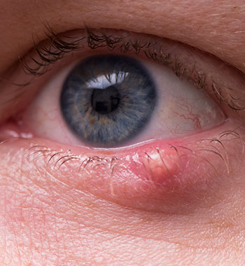
It has earned a reputation as “the great masquerader” of eyelid disease because it so convincingly mimics far more common, entirely benign conditions, chalazion and chronic blepharoconjunctivitis chief among them.
This mimicry, combined with a real tendency toward pagetoid intraepithelial spread that extends well beyond what is visible on the surface, is what makes delayed diagnosis so consequential and so unfortunately common.
Why It’s So Often Missed
A substantial proportion of patients are initially treated for presumed recurrent chalazion or chronic blepharitis, sometimes for many months, before the correct diagnosis is finally made, and this diagnostic delay is one of the most consistently cited factors associated with worse outcomes in sebaceous carcinoma specifically.
Any “chalazion” that recurs at the same site after treatment, or that fails to respond as expected to standard incision and curettage, deserves genuine suspicion and, ultimately, biopsy rather than repeated conservative treatment.
Building this specific threshold into routine practice — biopsying a second recurrence rather than simply repeating incision and curettage a third time — is genuinely one of the more consequential habits an ophthalmologist can adopt, given how directly diagnostic delay correlates with worse outcomes in this particular tumor.
Risk Factors
- Older age, with peak incidence in the seventh and eighth decades
- Prior radiation exposure to the eyelid or periocular region
- Muir-Torre syndrome — a genetic condition associated with sebaceous neoplasms (including sebaceous carcinoma) and an increased risk of internal malignancy, particularly colorectal cancer; recognizing sebaceous carcinoma can therefore be a signal to consider broader genetic and systemic cancer screening in the right clinical context
- Higher reported incidence in some Asian populations relative to Western populations
All-fit Slit-Lamp Adapter
Record and share exactly what you see at the slit lamp. One adapter fits any slit lamp or surgical microscope — and any smartphone.
From Choroida — the team behind this siteClinical Presentation
- A firm, painless, yellowish nodule, most often on the upper eyelid (where meibomian gland density is greater), frequently mistaken for a chalazion
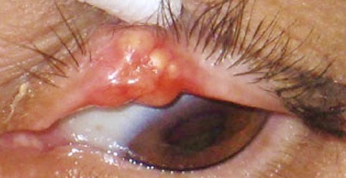
- Diffuse thickening and induration of the eyelid margin in the pagetoid spread pattern, sometimes presenting as a chronic unilateral blepharoconjunctivitis rather than a discrete nodule
- Loss of eyelashes (madarosis) in the affected area — a genuinely useful clinical clue, since ordinary chalazion does not typically cause lash loss
- Recurrence at the same site after treatment presumed to be for a benign chalazion
- Yellow discoloration of the tarsal conjunctiva on lid eversion, reflecting the underlying lipid-rich tumor
Differential Diagnosis
- Chalazion — the most common and most consequential misdiagnosis; distinguishing features favoring carcinoma include recurrence at the same site, madarosis, diffuse lid thickening, and older patient age
- Chronic blepharitis or blepharoconjunctivitis — the pagetoid spread pattern can closely mimic this, particularly when unilateral and treatment-resistant
- Basal cell carcinoma — the most common eyelid malignancy overall, but generally slower growing, with a more classic pearly, rolled-border appearance and lower metastatic potential than sebaceous carcinoma
- Squamous cell carcinoma — can appear similarly, definitively distinguished on histopathology
Diagnostic Evaluation
Any suspicious eyelid lesion — a “chalazion” recurring at the same site, an atypical or treatment-resistant chronic blepharoconjunctivitis, or a lesion with associated madarosis — warrants biopsy rather than continued conservative management.
Because the tumor can spread in a pagetoid (intraepithelial, multifocal) pattern well beyond the visible extent of the lesion, map biopsies of the conjunctiva are sometimes used to define the true extent of involvement before definitive surgical planning, since relying on the visible lesion alone can significantly underestimate how much tissue is actually involved.
Management
Wide surgical excision with margin control — Mohs micrographic surgery or frozen-section-controlled excision — is the standard of care, chosen specifically because of the tumor’s tendency toward multifocal, pagetoid spread that simple excision with standard margins can under-treat.
Given the risk of regional lymphatic spread, sentinel lymph node biopsy is considered in cases with high-risk features (larger tumor size, orbital invasion, or other adverse histologic characteristics), following similar staging principles used for other cutaneous malignancies with meaningful metastatic potential.
Orbital exenteration is reserved for cases with extensive orbital invasion not amenable to more conservative surgery, a genuinely disfiguring intervention that underscores how much is at stake with delayed diagnosis and treatment of this tumor.
Radiation therapy has a role for patients who are not surgical candidates or as an adjunct in select cases, though surgery with adequate margin control remains the preferred primary treatment given the tumor’s propensity for local recurrence when margins are inadequately assessed.
Prognosis
Prognosis correlates strongly with how early the diagnosis is made.
Tumors caught while still small and confined carry a substantially better prognosis than those diagnosed after prolonged mistreatment as a benign condition, reinforcing why a low threshold for biopsying an atypical or recurrent eyelid lesion is so clinically important.
Regional and distant metastasis, though less common than local recurrence, carries a significantly worse prognosis and underscores the seriousness of this diagnosis relative to the far more common, entirely benign conditions it so often resembles.
Long-term follow-up after treatment, including monitoring for both local recurrence and regional lymphatic spread, remains important for years, since this tumor’s capacity for late recurrence means a single clear follow-up visit shortly after surgery does not fully establish that the disease has been eradicated.


Document what you see
Two smartphone imaging tools built for everyday clinic use — one for the slit lamp, one for the fundus.
From Choroida — the team behind this siteReferences
- Shields JA, Demirci H, Marr BP, et al. Sebaceous carcinoma of the eyelids: personal experience with 60 cases. Ophthalmology.
- Kass LG, Hornblass A. Sebaceous carcinoma of the ocular adnexa. Survey of Ophthalmology.
- American Academy of Ophthalmology. Basic and Clinical Science Course, Section 7: Orbit, Eyelids, and Lacrimal System.
Test yourself
A few questions straight from this article.
-
From which structures does eyelid sebaceous carcinoma arise?
This uncommon but dangerous malignancy arises from the sebaceous glands of the ocular adnexa: the meibomian glands, the glands of Zeis, or the sebaceous glands of the caruncle. -
In which decades does eyelid sebaceous carcinoma peak in incidence?
Older age is a principal risk factor, with peak incidence in the seventh and eighth decades. Prior periocular radiation is another recognised risk factor. -
Which internal malignancy risk does Muir-Torre syndrome carry alongside sebaceous neoplasms?
Muir-Torre syndrome links sebaceous neoplasms with increased internal malignancy risk, particularly colorectal cancer, so the eyelid tumour can be the signal to consider broader genetic and systemic screening. -
Which eyelid sign favours sebaceous carcinoma over an ordinary chalazion?
Madarosis is a genuinely useful clue, since an ordinary chalazion does not typically cause lash loss. Diffuse lid thickening and older patient age likewise favour carcinoma. -
What does yellow discoloration of the tarsal conjunctiva on lid eversion reflect in sebaceous carcinoma?
Everting the lid can reveal yellow discoloration of the tarsal conjunctiva, a direct reflection of the lipid-rich sebaceous tumour beneath it. -
Why are conjunctival map biopsies sometimes performed in sebaceous carcinoma?
The tumour spreads in a pagetoid, intraepithelial, multifocal pattern well beyond the visible lesion, so relying on what is visible alone can significantly underestimate how much tissue is involved. -
What should be done with a chalazion recurring at the same site after treatment?
Diagnostic delay is the factor most consistently linked to worse outcomes in sebaceous carcinoma. Biopsying a second recurrence rather than repeating incision and curettage is a consequential habit to build in. -
Why is margin-controlled excision the standard of care for eyelid sebaceous carcinoma?
Mohs micrographic surgery or frozen-section-controlled excision is chosen specifically because the tumour's multifocal, pagetoid spread means simple excision with standard margins can leave disease behind. -
When is sentinel lymph node biopsy considered in eyelid sebaceous carcinoma?
Given the risk of regional lymphatic spread, sentinel node biopsy is considered for larger tumours, orbital invasion, or other adverse histologic characteristics, following staging principles used for other cutaneous malignancies. -
Why is prolonged follow-up needed after treating eyelid sebaceous carcinoma?
Monitoring for both local recurrence and regional lymphatic spread matters for years, because a single clear visit shortly after surgery does not establish that the disease has been eradicated.
