
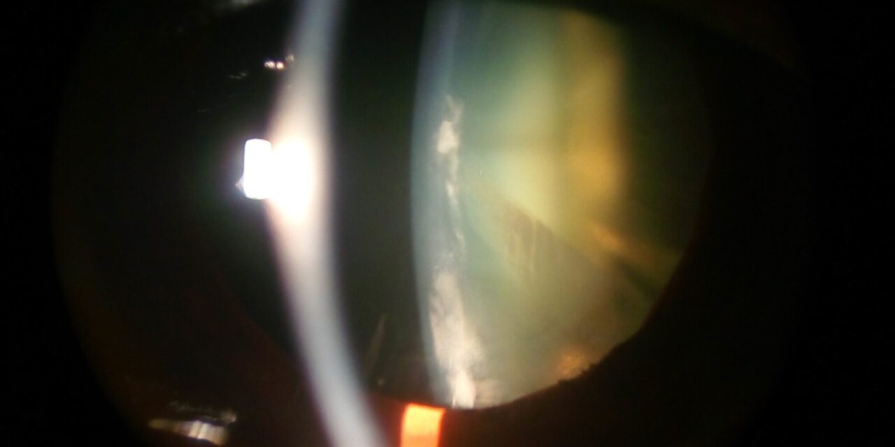
A membranous cataract is what remains when the contents of the lens have been reabsorbed.
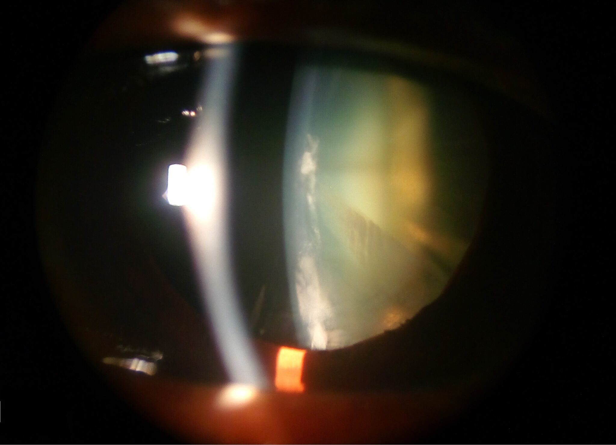
The capsule collapses, the anterior and posterior layers fuse, and a thin, white or gray, sometimes calcified, sheet is left in the pupillary area.
It is an end-stage lens, and the visual axis is blocked by a dense membrane instead of a swollen, cloudy lens.
How it develops
The lens is made of a clear capsule filled with fibers.
If the capsule is breached, the lens substance can be absorbed by macrophages, as in a child after penetrating injury.
In congenital cataract, particularly after intrauterine infection or in certain syndromes, the lens fibers may be abnormal, and over time they degenerate and resorb.
The remaining capsule and fibrous tissue form a flat membrane.
Settings in which it occurs
- Congenital rubella syndrome, in which pearly cataract with residual membrane is common (see congenital rubella syndrome)
- Penetrating or blunt trauma in children, with capsule rupture and spontaneous absorption of lens cortex
- Lowe oculocerebrorenal syndrome and some other metabolic or genetic conditions
- Chronic uveitis and long-standing inflammation
- Long-standing congenital cataract left untreated
- Incomplete lens removal in earlier surgery, with a residual opaque membrane
All-fit Slit-Lamp Adapter
Record and share exactly what you see at the slit lamp. One adapter fits any slit lamp or surgical microscope — and any smartphone.
From Choroida — the team behind this siteWhat the examiner sees
The pupil is filled by a white or gray membrane, sometimes with a few strands attached to the iris.
The anterior chamber may be deep because the lens volume has shrunk, and the iris may tremble (iridodonesis).
Details of the fundus are not visible.
There may be calcium deposits, and posterior synechiae are common.
In a young child, the eye may show nystagmus or poor fixation if the condition is bilateral, and strabismus if unilateral.
Work-up
- B-scan ultrasonography to assess the posterior segment, since a retinal detachment, microphthalmia, or persistent fetal vasculature may be present
- Biometry to plan intraocular lens implantation, if appropriate
- Investigation for the cause, including TORCH serology, metabolic screening, and a systemic review in children
- Slit-lamp or handheld examination in the clinic, or under anesthesia in young children
Treatment
Surgery is the only effective treatment.
The membrane is removed or opened with a vitreous cutter, scissors, or a capsulotomy, with an anterior vitrectomy to prevent re-opacification, and intraocular lens implantation is considered depending on age and eye size.
In rubella cataract, the surgeon should be aware that live virus can persist in the lens, and the eye may be inflamed after surgery, so inflammation control is important.
In children, amblyopia is the major threat, so surgery should be done early, followed by optical correction and patching or other amblyopia therapy (see pediatric cataract surgery).
Assessing the eye before surgery
A membranous cataract may hide other problems, so the examination should be thorough. Measure the intraocular pressure, check for a relative afferent pupillary defect, and assess whether the eye follows light and objects. Ultrasound helps to determine the axial length, the status of the vitreous, and the position of the retina. In children, an examination under anesthesia may be needed to complete the assessment.
Surgical approach
The membrane is often tough and adherent, and a vitrectomy cutter or scissors may be needed to open it. The aim is to create a clear central opening, to remove the membrane as completely as is safe, and to perform an anterior vitrectomy to reduce the risk of opacification. Intraocular lens implantation may be postponed in an eye that is small, inflamed, or at high risk for complications.
Aftercare
Inflammation is controlled with topical steroids and cycloplegics. Optical correction with contact lenses or spectacles is started early, and patching or other amblyopia treatment follows the usual principles. Long-term follow-up is needed for glaucoma, which can appear years after surgery in children with congenital cataract.
A note on glaucoma and amblyopia
Children with congenital or traumatic membranous cataract are at risk for glaucoma after surgery, sometimes many years later, and need lifelong pressure monitoring. Amblyopia is the greatest threat to vision in children. Early surgery, correction of aphakia, and patching or other amblyopia therapy are the foundations of a good outcome, and families need strong support to maintain treatment over years.
Differential diagnosis
A membranous lens can be confused with a retrolental mass, persistent fetal vasculature, a dense posterior capsule opacity after earlier surgery, or a fibrous plaque from inflammation. Ultrasound and careful examination under anesthesia help to separate them, and a tumor such as retinoblastoma must always be excluded when the fundus cannot be seen in a child with leukocoria.
Prognosis
Outcomes depend on the age at surgery, the presence of other eye anomalies, and the quality of amblyopia treatment.
Eyes with microphthalmia, glaucoma, or retinal disease have a more guarded outlook.


Document what you see
Two smartphone imaging tools built for everyday clinic use — one for the slit lamp, one for the fundus.
From Choroida — the team behind this siteReferences
- Givens KT, Lee DA, Jones T, Ilstrup DM. Congenital rubella syndrome: ophthalmic manifestations and associated systemic disorders. Br J Ophthalmol. 1993;77:358-363.
- Lambert SR, Drack AV. Infantile cataracts. Surv Ophthalmol. 1996;40:427-458.
- Wilson ME, Trivedi RH, Pandey SK. Pediatric Cataract Surgery: Techniques, Complications, and Management. Philadelphia: Lippincott Williams & Wilkins; 2005.
- Duke-Elder S. System of Ophthalmology. Vol XI: Diseases of the Lens and Vitreous; Glaucoma and Hypotony. London: Kimpton; 1969.
