
CASE REPORT
A 48-year-old male with a history of hypertension and hypercholesterolemia presented with a two-week history of sudden, painless visual disturbances in his right eye, including floating spots and flashing lights in the nasal visual field.
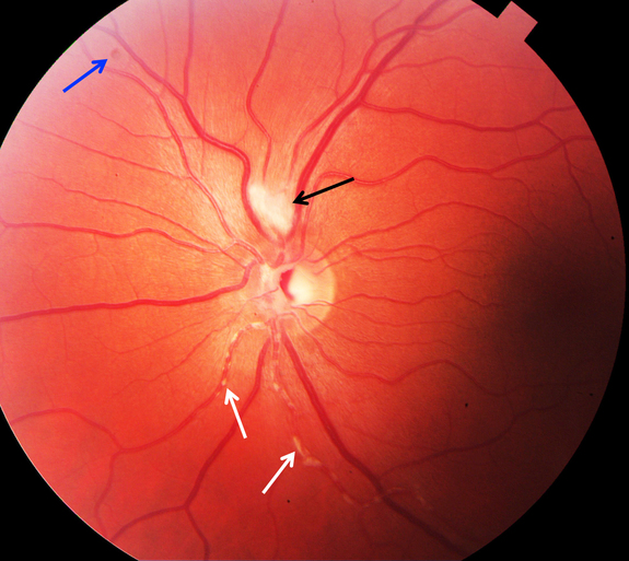
On examination, his right eye had a visual acuity of 20/30, while the left eye was 20/20. Fundus examination of the right eye revealed yellowish-white plaques along arterioles without macular involvement, alongside a relative afferent pupillary defect (RAPD).
No intraocular inflammation or other abnormalities were noted. Diagnostic workup included optical coherence tomography (OCT), fluorescein angiography (FA) indicating arterial wall staining and dye leakage at plaque sites, and blood tests for autoimmune markers (ANA and antiphospholipid antibodies), all of which were negative. The diagnosis of Kyrieleis plaques was confirmed.
Kyrieleis Plaques entity
Kyrieleis plaques are also referred to as “Kyrieleis vasculitis”, “Kyrieleis arteriolitis”, “nodular periarteritis”, “segmental retinal periarteritis” or “segmental retinal arteritis” represent a rare fundus examination finding, first described in an eye with presumed tuberculous uveitis by Werner Kyrieleis, in 1933.
They appear as multiple, segmental yellowish-white lesions within the retinal arteries, in a beaded pattern reported both in infectious and non-infectious inflammatory posterior uveitis. These plaques are usually reversible.
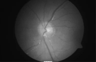
The etiology of Kyrieleis plaques is unknown but they always reflect severe intra-ocular inflammation. They have been typically associated with infectious posterior uveitis, and Toxoplasma gondii is the most frequently reported cause.
Treponema pallidum, Mycobacterium tuberculosis, Varicella-Zoster Virus, Cytomegalovirus, Herpes Simplex Virus- 1 and 2, and Rickettsia conorii are other common agents.
Kyrieleis arteritis in association with Behçet disease has also been described. Kyrieleis plaques are also described as a clinical feature of Brolucizumab-Associated Retinal Vasculitis and Intraocular Inflammation.
The pathophysiology of these plaques is not completely known, and there is still a lot to discover about the composition of the whitish deposits and their exact location within the arteries.
Various hypotheses have been proposed, though. Griffin and Bodian, in 1959, speculated that the plaques represented exudates migration from an adjacent retinochoroiditis focus to the periarterial sheaths.
In 1971, Orzalesi and Ricciardi hypothesized that these deposits constituted cellular and inflammatory material within the arterial walls. Wise proposed that they were arteriosclerosis lesions.
Recently, a study based on multimodal retinal imaging, suggested that the plaques do not represent periarterial or endoluminal injury, but the involvement of the arterial endothelium.
Fundus Explorer Pro
Photograph the retinal findings described here with the phone already in your pocket — 22 D optics and built-in illumination in one handheld unit.
From Choroida — the team behind this siteDiagnosis of Kyrieleis Plaques
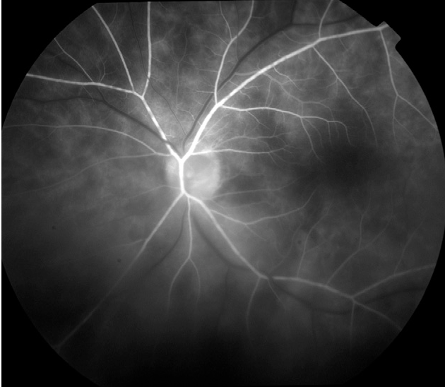
Like other forms of retinal vasculitis, the diagnosis of Kyrieleis is clinical, based on the fundoscopic examination, and supported by ancillary tests namely fluorescein angiography (FA), indocyanine green (ICG) angiography, fundus autofluorescence (FAF), and optical coherence tomography (OCT).
Fundoscopy
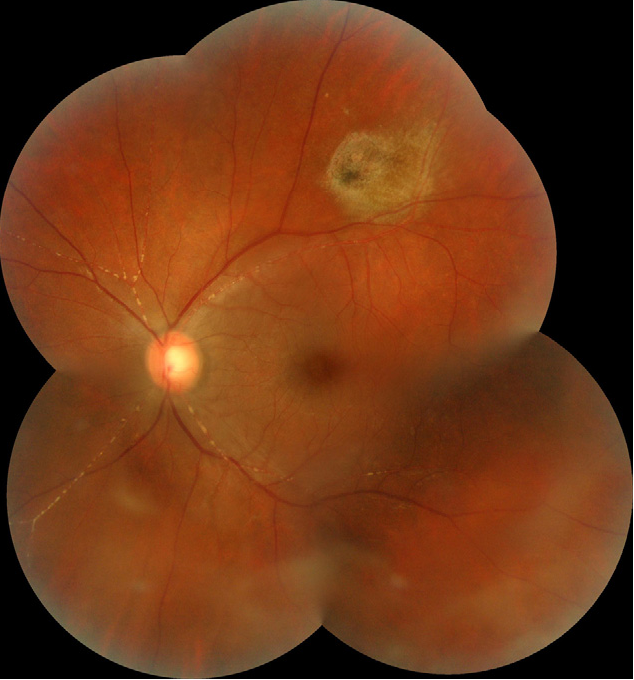
On the Smartphone Fundoscopy, Kyrieleis plaques appear as focal yellowish-white deposits, looking like atheromatous plaques, segmentally distributed along the retinal arteries.
As previously mentioned, these plaques always occur in association with intra-ocular inflammation, and typically, an active focus of chorioretinitis is present near the affected arteries.
Sometimes, the plaques can only be visible once the severe intraocular inflammation has resolved. Kyrieleis plaques involve exclusively the arterial and glisten and often look like calcific lesions.
Kyrieleis Plaques MANAGEMENT
The management of Kyrieleis arteritis depends on the underlying etiology. The control of intraocular inflammation is the primary goal.
Although associated with severe ocular inflammation, Kyrieleis plaques are a benign finding, that does not worsen the prognosis of the underlying uveitis.
Generally, with adequate treatment, there is a complete disappearance of the plaques (usually after the resolution of vitritis) without sequelae 5.
However, the plaques can persist long after the resolution of chorioretinitis and discontinuation of therapy.


Document what you see
Two smartphone imaging tools built for everyday clinic use — one for the slit lamp, one for the fundus.
From Choroida — the team behind this siteREFERENCES
- Kyrieleis W. [Discontinuous reversible arteriopathy in uveitis]. Albrecht Von Graefes Arch Ophthalmol. 1950;150(5-6):600-13.
- Francés-Muñoz E, Gallego-Pinazo R, López-Lizcano R, García-Delpech S, Mullor JL, Díaz-Llopis M. Kyrieleis’ vasculitis in acute retinal necrosis. Clin Ophthalmol. 2010;4:837-8.
- Goel N, Sawhney A. Kyrieleis plaques associated with Herpes Simplex Virus type 1 acute retinal necrosis. Saudi J Ophthalmol. 2016;30(2):144-7.
- Pichi F, Veronese C, Lembo A, Invernizzi A, Mantovani A, Herbort CP, et al. New appraisals of Kyrieleis plaques: a multimodal imaging study. Br J Ophthalmol. 2017;101(3):316-21.
- Mahjoub A, Ben Abdesslem N, Zaafrane N, Sellem I, Sahraoui F, Nouri H, Hadj RB, Ben Alaya A, Krifa F, Hachemi M. Kyrieleis arteritis associated with toxoplasmic retinochoroiditis: A case report. Ann Med Surg (Lond). 2022 May 17;78:103802.
- Chazalon E, Conrath J, Ridings B, Matonti F. [Kyrieleis arteritis: report of two cases and literature review]. J Fr Ophtalmol. 2013;36(3):191-6.
Test yourself
A few questions straight from this article.
-
How do Kyrieleis plaques appear on fundus examination?
Kyrieleis plaques are multiple segmental yellowish-white lesions arranged in a beaded pattern, resembling atheromatous plaques. -
Along which retinal vessels are Kyrieleis plaques distributed?
The plaques lie within and along the retinal arteries, which is why the finding is also called segmental retinal arteritis. -
What does the presence of Kyrieleis plaques always indicate?
Although their cause is unknown, Kyrieleis plaques always reflect severe intraocular inflammation, reported in both infectious and non-infectious posterior uveitis. -
Which infectious agent is the most frequently reported cause of Kyrieleis plaques?
Kyrieleis plaques are typically associated with infectious posterior uveitis, and Toxoplasma gondii is the most frequently reported cause. -
Kyrieleis plaques have been described as a feature of intraocular inflammation after which intravitreal agent?
Kyrieleis plaques are described as a clinical feature of brolucizumab-associated retinal vasculitis and intraocular inflammation. -
What did a recent multimodal imaging study suggest about the site of Kyrieleis plaques?
Multimodal retinal imaging suggested the plaques represent involvement of the arterial endothelium rather than periarterial or endoluminal injury. -
How is the diagnosis of Kyrieleis plaques established?
As with other retinal vasculitides the diagnosis is clinical, made on fundoscopy and supported by fluorescein and ICG angiography, autofluorescence and OCT. -
What is typically found near the arteries affected by Kyrieleis plaques?
An active focus of chorioretinitis is typically present close to the arteries bearing the plaques, reflecting the associated intraocular inflammation. -
What is the primary goal when managing Kyrieleis arteritis?
Management is directed at the underlying cause, with control of the intraocular inflammation as the primary goal. -
How do Kyrieleis plaques affect the prognosis of the underlying uveitis?
Despite occurring with severe ocular inflammation, the plaques are a benign finding that does not worsen the prognosis of the underlying uveitis.
