
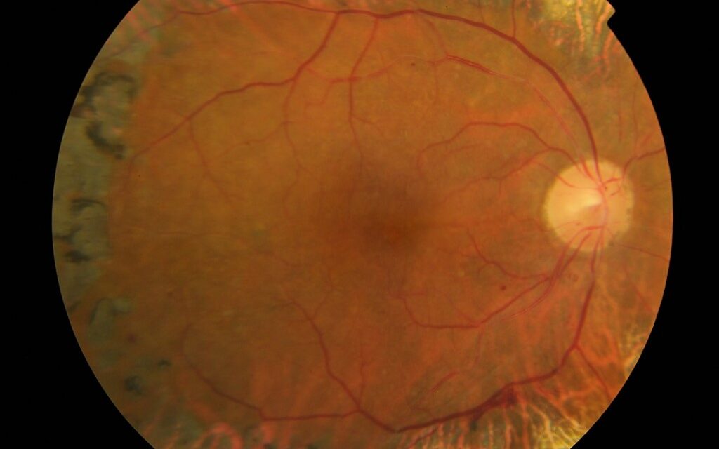
Vitreoretinal traction syndrome, more commonly referred to today as vitreomacular traction, represents a mechanical problem with a genuinely mechanical solution, and understanding its relationship to the normal process of vitreous separation from the retina explains both why it occurs in some eyes and not others, and why treatment options range from simple observation to enzymatic dissolution to surgery.
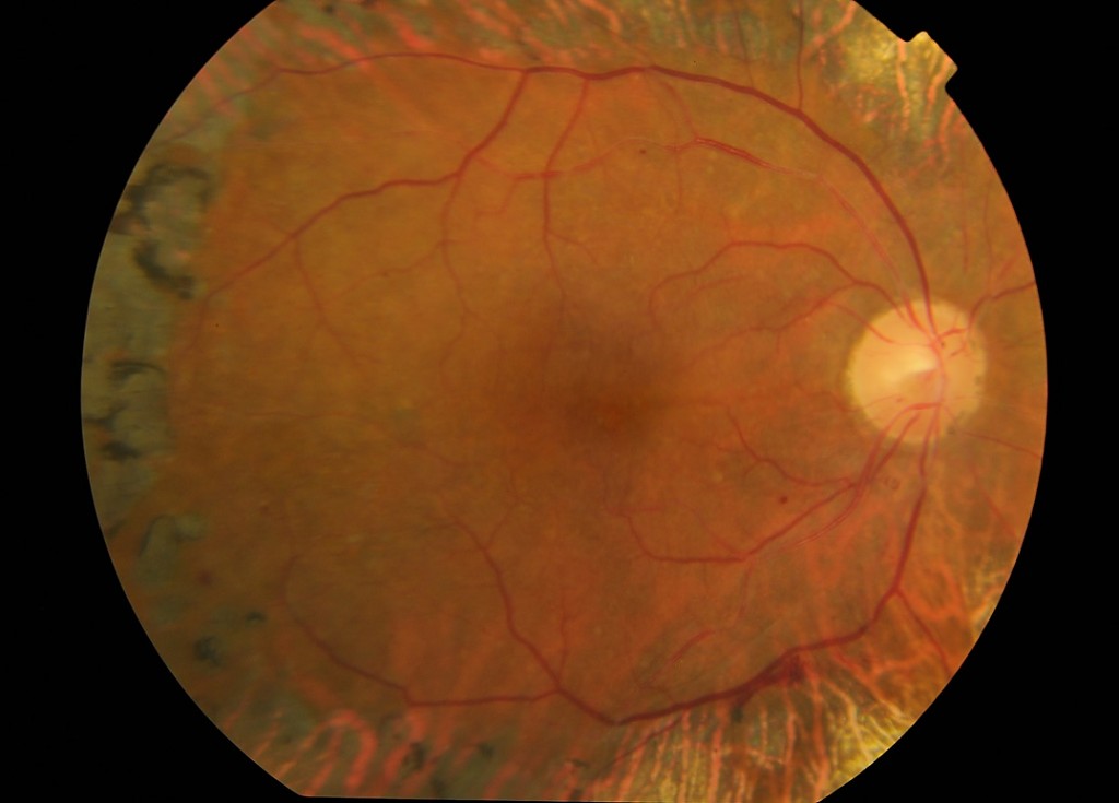
A Failure of Complete, Clean Vitreous Separation
As part of the normal aging process, the vitreous gel gradually liquefies and separates from the retinal surface, a process called posterior vitreous detachment, and in most eyes this separation proceeds cleanly and completely without consequence.
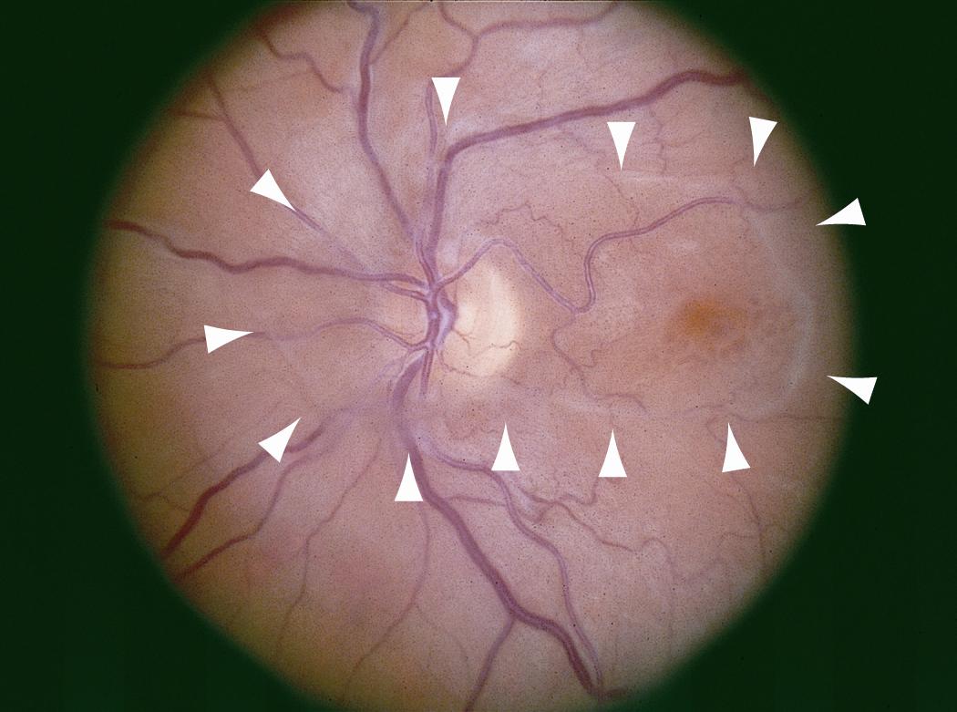
Vitreomacular traction occurs when this separation is incomplete, leaving a persistent area of vitreous attachment specifically at the macula while the surrounding vitreous has otherwise separated, and this residual, focal attachment exerts anteroposterior and tangential traction on the macula, capable of producing structural distortion and associated visual symptoms.
Clinical Features
- Metamorphopsia (visual distortion) and, depending on the severity and distribution of the traction, varying degrees of central vision reduction are the typical presenting symptoms
- On optical coherence tomography, a hallmark finding, persistent vitreous attachment to the fovea with associated distortion of the normal foveal contour, sometimes with an associated cystic change, subretinal fluid, or, in more severe cases, an associated macular hole
Fundus Explorer Pro
Photograph the retinal findings described here with the phone already in your pocket — 22 D optics and built-in illumination in one handheld unit.
From Choroida — the team behind this siteClassification by Adhesion Width
Vitreomacular traction is often characterized based on the width of the persistent vitreomacular adhesion, with focal adhesion (generally defined as less than 1500 micrometers) more likely to respond favorably to pharmacologic or spontaneous resolution, while broad adhesion is less likely to resolve without surgical intervention, a distinction with direct, practical treatment-selection implications.
Diagnostic Evaluation
- Optical coherence tomography is the central diagnostic tool, both confirming the diagnosis and characterizing the width and configuration of the vitreomacular adhesion, along with any associated structural macular changes, information that directly informs treatment planning
- Visual acuity and, when relevant, an Amsler grid assessment of metamorphopsia help correlate structural findings with functional visual impact
- Careful examination to identify any associated pathology, including full-thickness or lamellar macular hole, epiretinal membrane, or other coexisting vitreomacular interface abnormality
Management
Observation
Mild cases, particularly with minimal symptoms and a favorable, focal adhesion pattern, can be observed, since spontaneous release of the vitreomacular attachment occurs in a meaningful proportion of these more favorable cases over time.
Enzymatic Vitreolysis
Intravitreal ocriplasmin, an enzymatic agent capable of dissolving the vitreomacular adhesion, is an option for appropriately selected cases, particularly those with focal adhesion and without significant associated epiretinal membrane, since epiretinal membrane presence reduces the likelihood of successful enzymatic release.
Pars Plana Vitrectomy
Surgical vitrectomy, directly and mechanically separating the vitreous from the macula, is used for cases not suited to or not responsive to enzymatic treatment, particularly those with broad adhesion, significant associated epiretinal membrane, or more severe structural complications such as an associated macular hole (see vitreomacular adhesion for a related discussion of this vitreous-macular interface spectrum before it has progressed to symptomatic traction).
Symptoms patients report
Patients with vitreomacular traction may describe distorted or wavy lines, a blind spot, difficulty reading, and objects that look smaller or larger than normal. These symptoms may fluctuate. Examination with an Amsler grid shows distortion, and OCT shows the traction. Because the symptoms are subtle, many patients wait before seeking help.
Options in more detail
Ocriplasmin was used for focal adhesion, but it may cause temporary visual symptoms and, in rare cases, changes in the ellipsoid zone, so patients were selected carefully. Vitrectomy removes the traction directly, often with peeling of epiretinal membrane or the internal limiting membrane, and with a gas bubble if there is a hole. After surgery, the patient may need to position the head, and cataract formation is a common long-term effect.
What to tell patients
Explain that observation is reasonable for mild cases, that treatment aims to stop the traction before it damages the retina, and that vision often improves but may not return to normal. Warn about the symptoms of retinal detachment after surgery.
Prognosis
Visual outcomes after successful release of vitreomacular traction, whether through observation-associated spontaneous release, enzymatic vitreolysis, or surgery, are generally favorable, particularly when treatment occurs before prolonged, severe traction has produced more significant, potentially less reversible structural macular damage.
Appropriate treatment selection, guided by the width of adhesion and presence of associated pathology identified on optical coherence tomography, supports the best balance between avoiding unnecessary intervention in cases likely to resolve spontaneously and providing timely treatment for cases unlikely to improve without it.


Document what you see
Two smartphone imaging tools built for everyday clinic use — one for the slit lamp, one for the fundus.
From Choroida — the team behind this siteReferences
- Johnson MW. Posterior vitreous detachment: evolution and complications of its early stages. Am J Ophthalmol. 2010;149:371-382.e1.
- Duker JS, Kaiser PK, Binder S, et al. The International Vitreomacular Traction Study Group classification of vitreomacular adhesion, traction, and macular hole. Ophthalmology. 2013;120:2611-2619.
- Stalmans P, Benz MS, Gandorfer A, et al. Enzymatic vitreolysis with ocriplasmin for vitreomacular traction and macular holes. N Engl J Med. 2012;367:606-615.
- John VJ, Flynn HW Jr, Smiddy WE, et al. Clinical course of vitreomacular adhesion managed by initial observation. Retina. 2014;34:442-446.
