
Giant fornix syndrome is a diagnosis that depends entirely on a single examination maneuver most clinicians don’t routinely perform: deliberately everting and exploring the depth of the superior conjunctival fornix, since this condition’s defining feature, an abnormally deep, lax superior fornix serving as a hidden reservoir for chronic bacterial infection, is invisible without specifically looking for it.
The Underlying Mechanism
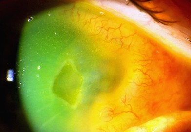
This condition occurs predominantly in elderly patients and is characterized by an abnormally deep and lax superior conjunctival fornix, which serves as a reservoir where bacterial biofilm, particularly involving Staphylococcus aureus, can accumulate and persist, driving a chronic, low-grade, often relapsing conjunctivitis that proves stubbornly resistant to standard, short-course topical antibiotic treatment.
Standard treatment courses fail repeatedly in this condition specifically because they do not address the anatomic reservoir itself, the deep, lax fornix harboring the persistent biofilm, meaning the bacterial burden simply re-establishes itself once a typical, time-limited antibiotic course ends.
Clinical Presentation
- Chronic, often relapsing, mucopurulent conjunctivitis, typically affecting older patients and often present for a prolonged period, sometimes months to years, before the correct underlying cause is identified
- Recurrence or incomplete resolution despite multiple courses of standard topical antibiotic treatment, a pattern that should specifically raise suspicion for this diagnosis
- On examination with deliberate eversion and exploration of the superior fornix, an abnormally deep, lax fornix is identified, often with visible mucopurulent discharge or debris pooled within it
- Associated floppy eyelid syndrome and other age-related eyelid laxity changes are sometimes present concurrently, reflecting shared underlying connective tissue laxity in this patient population
All-fit Slit-Lamp Adapter
Record and share exactly what you see at the slit lamp. One adapter fits any slit lamp or surgical microscope — and any smartphone.
From Choroida — the team behind this siteWhy This Diagnosis Is So Often Missed
A routine external examination, without deliberate, deep eversion of the superior fornix specifically to assess its depth and contents, will not reveal this condition, and a patient can go through multiple courses of standard antibiotic treatment for presumed ordinary bacterial or chronic conjunctivitis without the underlying anatomic reservoir ever being identified, which is precisely why persistent, recurrent conjunctivitis unresponsive to standard treatment in an older patient should specifically prompt this more deliberate examination technique.
Diagnostic Evaluation
- Deliberate eversion and exploration of the superior conjunctival fornix, using a cotton-tip applicator or similar instrument to assess fornix depth and directly visualize any pooled discharge or debris, the essential examination step for reaching this diagnosis
- Conjunctival culture from the fornix reservoir, often revealing heavy growth of Staphylococcus aureus, supporting both the diagnosis and antibiotic selection
- A history of chronic or recurrent conjunctivitis resistant to standard, short-course topical antibiotic treatment, which should prompt specific consideration of this diagnosis and the deeper fornix examination needed to confirm it
Management
Mechanical Debridement
Mechanical cleaning and debridement of the fornix reservoir, removing accumulated biofilm and debris, is an important part of treatment, directly addressing the physical reservoir that perpetuates the chronic infection, and this is often repeated over the course of treatment rather than performed only once.
Combined Antibiotic and Anti-Inflammatory Treatment
A combination of topical antibiotic treatment, often intensive and prolonged compared with standard conjunctivitis management, together with topical corticosteroid treatment to address the associated inflammatory component, is generally used, sometimes described as a “pulse” regimen with periodic intensive treatment courses.
Addressing Fornix Laxity
For refractory cases, surgical procedures aimed at reducing the excessive fornix depth and laxity have been described, directly addressing the underlying anatomic predisposition in patients where medical management alone proves insufficient to achieve durable control.
How to examine the fornix
Instill a drop of topical anesthetic, ask the patient to look down, and evert the upper lid fully, using a cotton-tipped applicator to push the fornix open. Look for pooled mucus, pus, and debris in the depth of the fornix, and compare with the other side. Take a swab from the depth of the fornix for culture before cleaning. Many patients with floppy eyelids and lax tissues will have a particularly deep fornix.
Practical treatment tips
Clean the fornix with saline and a cotton applicator at the visit, and teach the patient to use warm compresses and lid hygiene. Treat the infection with antibiotics chosen by culture, and add a steroid cautiously for inflammation. Review frequently, and consider surgery if the problem returns despite treatment. Explain that the disease tends to relapse if the reservoir is not cleared.
Prognosis
Giant fornix syndrome, once correctly identified through the deliberate fornix examination this diagnosis requires, generally responds well to a combination of mechanical debridement and appropriately intensive, sustained antibiotic and anti-inflammatory treatment.
The central challenge in this condition is diagnostic rather than therapeutic, and recognizing the pattern, chronic, recurrent conjunctivitis resistant to standard treatment in an older patient, that should prompt the specific fornix examination needed to reach this diagnosis is the genuinely important clinical skill this condition requires.


Document what you see
Two smartphone imaging tools built for everyday clinic use — one for the slit lamp, one for the fundus.
From Choroida — the team behind this siteReferences
- Rose GE. The giant fornix syndrome: an unrecognized cause of chronic, relapsing, grossly purulent conjunctivitis. Ophthalmology. 2004;111:1539-1545.
- Rose GE, Lavy FJ. Giant fornix syndrome: a case series. Ophthalmology. 2005;112:1539-1545.
- Vinciguerra P, Rosetta P, Legrottaglie EF, et al. Giant fornix syndrome: a review. Surv Ophthalmol. 2018;63:531-537.
- Sridhar U, Tripathy K. Giant fornix syndrome. StatPearls. Treasure Island (FL): StatPearls Publishing; 2023.
