
Case study
A 47-year-old female presented to the ophthalmology clinic with a two-year history of progressively worsening ocular symptoms, including redness, irritation, and blurred vision.
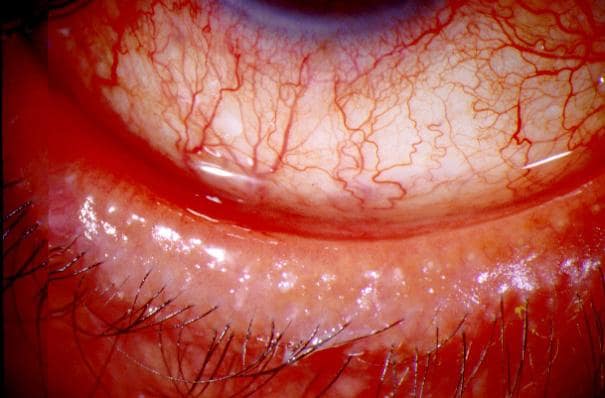
She reported a burning sensation, foreign body sensation, and increased sensitivity to light. The patient had a known history of cutaneous rosacea, diagnosed ten years prior, for which she was intermittently treated with topical metronidazole and oral doxycycline.
Her medical history included mild hypertension managed with lisinopril and seasonal allergies treated with over-the-counter antihistamines.
Her family history included her mother having rosacea and dry eye syndrome. On examination, the patient’s visual acuity was 20/25 in both eyes.
Slit-lamp examination revealed telangiectasia of the eyelid margins, posterior blepharitis with visible meibomian gland dysfunction, moderate conjunctival hyperemia, and punctate epithelial erosions on the cornea.
The patient’s skin showed typical signs of rosacea, including erythema and papules on the cheeks and nose. A diagnosis of ocular rosacea was made based on the clinical presentation and examination findings,
Ocular Rosacea entity
Ocular rosacea is a chronic inflammatory condition affecting the eyes, often in conjunction with the skin disorder rosacea.
This condition can cause significant discomfort and may lead to serious complications, including vision loss, if not properly managed.
Understanding the pathogenesis, epidemiology, clinical features, diagnosis, differential diagnosis, and management of ocular rosacea is essential for effective treatment and patient care.
All-fit Slit-Lamp Adapter
Record and share exactly what you see at the slit lamp. One adapter fits any slit lamp or surgical microscope — and any smartphone.
From Choroida — the team behind this sitePathogenesis
The exact cause of ocular rosacea is not fully understood, but it likely involves a combination of genetic predisposition, immune system dysfunction, and environmental factors.
The inflammation primarily affects the eyelids (blepharitis), conjunctiva, and sometimes the cornea. Several mechanisms have been proposed to explain the pathogenesis of ocular rosacea:
- Immune System Dysfunction: Aberrant immune responses may lead to chronic inflammation in ocular tissues. An imbalance between pro-inflammatory and anti-inflammatory cytokines can result in persistent inflammation and tissue damage.
- Microbial Factors: Demodex mites and bacteria, such as Staphylococcus species, are often found in higher numbers on the eyelashes and eyelids of patients with rosacea. These microorganisms may trigger or exacerbate inflammation.
- Vascular Abnormalities: Telangiectasia, or dilated blood vessels, are common in rosacea and may contribute to ocular symptoms by increasing vascular permeability and promoting inflammation.
- Genetic Factors: Family history and genetic predisposition play roles in the development of rosacea, including its ocular manifestations.
Epidemiology
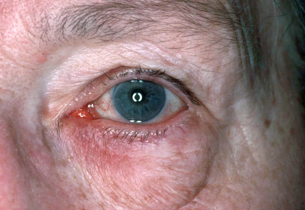
Ocular rosacea affects a significant proportion of individuals with rosacea, with estimates ranging from 6% to 58%. It typically manifests in adults between the ages of 30 and 50, with a higher prevalence among fair-skinned individuals.
Both genders are affected, although women are more frequently diagnosed with cutaneous rosacea, while men tend to present with more severe ocular symptoms.
Clinical Features
Patients with ocular rosacea may experience a variety of symptoms, including:
- Redness and Swelling of the Eyelids: Inflammation of the eyelids (blepharitis) can cause noticeable redness and swelling.
- Grittiness or Foreign Body Sensation: Patients often feel as though there is something in their eyes.
- Watery or Bloodshot Eyes: Increased tear production and visible redness of the eyes are common.
- Burning or Stinging Sensation: Inflammation can cause discomfort and a burning or stinging sensation in the eyes.
- Light Sensitivity (Photophobia): Patients may become more sensitive to light.
- Blurred Vision: Severe inflammation can affect the cornea, leading to blurred vision.
Ocular Rosacea Diagnosis
Diagnosis is primarily clinical, based on a thorough examination of the eyes and eyelids. Key diagnostic signs include:
- Telangiectasia of the Eyelid Margins: Visible small blood vessels on the eyelid margins are a hallmark of ocular rosacea.
- Meibomian Gland Dysfunction: Dysfunction of these glands, which secrete oils that form part of the tear film, can lead to dry eyes.
- Conjunctival Hyperemia: Redness of the conjunctiva is commonly observed.
- Corneal Involvement: This can include punctate keratitis, corneal infiltrates, or, in severe cases, corneal ulceration.
Differential Diagnosis
Ocular rosacea should be differentiated from other ocular conditions that may present with similar symptoms:
- Allergic Conjunctivitis: Allergic reactions can cause redness, itching, and swelling similar to ocular rosacea.
- Chronic Blepharitis Unrelated to Rosacea: Inflammation of the eyelids can occur without the presence of rosacea.
- Dry Eye Syndrome: Reduced tear production can lead to similar symptoms of dryness and irritation.
- Infectious Conjunctivitis: Bacterial or viral infections can cause redness, discharge, and discomfort.
- Autoimmune Disorders: Conditions such as Sjogren’s syndrome or systemic lupus erythematosus can affect the eyes.
Management of Ocular Rosacea
Management of ocular rosacea aims to control symptoms, reduce inflammation, and prevent complications. A multidisciplinary approach, involving both dermatologists and ophthalmologists, is often necessary.
Eyelid Hygiene
- Regular Cleaning: Cleaning the eyelid margins with warm compresses and gentle scrubbing can help reduce inflammation and remove debris.
Lubrication
- Artificial Tears: These can alleviate dryness and irritation by providing additional moisture to the eyes.
Topical Antibiotics
- Metronidazole Gel or Erythromycin Ointment: These can be applied to the eyelids to reduce inflammation and bacterial load.
Oral Antibiotics
- Tetracyclines: Medications such as doxycycline or minocycline are often prescribed for their anti-inflammatory properties. These are typically used in low doses over a prolonged period.
Immunomodulatory Agents
- Cyclosporine Eye Drops: In severe cases, these medications may be considered to reduce inflammation.
Lifestyle Modifications
- Avoiding Triggers: Avoidance of known triggers such as spicy foods, hot beverages, alcohol, and extreme temperatures can help manage symptoms.

Prognosis
With appropriate management, most patients with ocular rosacea can achieve good control of their symptoms. The condition, however, is chronic and may require ongoing treatment and regular follow-up to prevent relapses.
Early intervention is crucial to prevent potential complications, such as corneal damage and vision loss.


Document what you see
Two smartphone imaging tools built for everyday clinic use — one for the slit lamp, one for the fundus.
From Choroida — the team behind this siteReferences
- Akpek, E.K., and B.G. Gottsch. “Immune defense at the ocular surface.” Eye. 2003.
- Vieira, A.C., and C.L. Mannis. “Ocular rosacea: common and commonly missed.” Journal of the American Academy of Dermatology. 2013.
- Lemp, M.A., and E. S. Foulks. “The definition and classification of dry eye disease.” Ocular Surface. 2007.
- McDonnell, P.J., and G.N. Patrinely. “Ocular rosacea.” Survey of Ophthalmology. 1987.
- Wilkin, J.K., et al. “Standard classification of rosacea: report of the National Rosacea Society Expert Committee on the Classification and Staging of Rosacea.” Journal of the American Academy of Dermatology. 2002.
Test yourself
A few questions straight from this article.
-
Ocular rosacea is best described as which kind of condition?
Ocular rosacea is a chronic inflammatory condition of the eyes that often accompanies cutaneous rosacea, and it can cause real discomfort and even vision loss if poorly managed. -
Which eyelid sign is regarded as a hallmark of ocular rosacea?
Visible small vessels along the eyelid margins are the hallmark of ocular rosacea, seen alongside meibomian gland dysfunction and conjunctival hyperaemia. -
What proportion of patients with rosacea are estimated to have ocular involvement?
Estimates of ocular involvement among rosacea patients range widely, from 6% to 58%, with onset typically between 30 and 50 years of age. -
Which microorganisms are found in higher numbers on the lids and lashes in rosacea?
Demodex mites and Staphylococcus species are often present in greater numbers on the lids and lashes of rosacea patients and may trigger or worsen the inflammation. -
How does meibomian gland dysfunction contribute to the symptoms of ocular rosacea?
The meibomian glands secrete the oils that form part of the tear film, so their dysfunction in ocular rosacea leads to dry eye and the associated grittiness and burning. -
Which corneal changes may occur in ocular rosacea?
Corneal involvement runs from punctate keratitis through corneal infiltrates to frank ulceration in severe disease, which is why early intervention matters. -
Which oral antibiotic class is used in ocular rosacea for its anti-inflammatory effect?
Tetracyclines, usually doxycycline or minocycline, are prescribed for their anti-inflammatory properties, typically at low dose over a prolonged period. -
Which topical immunomodulator may be considered for severe ocular rosacea?
Cyclosporine eye drops are the immunomodulatory option reserved for severe cases, added to lid hygiene, lubrication and topical or oral antibiotics. -
Which lifestyle triggers should patients with ocular rosacea be advised to avoid?
Avoiding known triggers such as spicy food, hot beverages, alcohol and extremes of temperature helps control symptoms alongside medical treatment. -
What is the expected course of ocular rosacea under appropriate management?
Most patients achieve good symptom control, but the condition is chronic and needs ongoing treatment and regular review to prevent relapse and corneal damage.
