
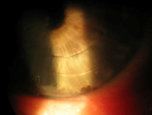
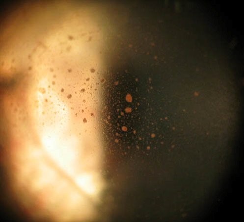
Shafer sign, the presence of pigmented cells scattered in the anterior vitreous, colloquially and memorably called “tobacco dust,” is one of the more valuable single findings in ophthalmic examination, because its presence carries a strong statistical association with an underlying retinal break, making it a finding that should prompt a specifically thorough, deliberate peripheral retinal examination rather than being noted and passed over.
Where the Pigment Comes From
The pigmented cells responsible for Shafer sign originate from the retinal pigment epithelium, and their presence in the vitreous means that retinal pigment epithelial cells have somehow gained access to the vitreous cavity, a event that, outside of specific alternative explanations, most often occurs at the site of a retinal break, where the neurosensory retina and underlying retinal pigment epithelium have been disrupted, allowing pigment cells to be liberated into the vitreous.
Clinical Appearance
- Fine, brown to gray, dust-like pigmented cells visible in the anterior vitreous on slit-lamp examination, most easily appreciated with a narrow beam and moderate to high magnification
- Often more concentrated in the anterior, retrolental vitreous, which is why specifically directing examination attention to this area, rather than only the central or posterior vitreous, improves detection
- Can range from sparse, subtle scattering to more dense, obvious pigment dispersion depending on the extent of the underlying retinal disruption
Fundus Explorer Pro
Photograph the retinal findings described here with the phone already in your pocket — 22 D optics and built-in illumination in one handheld unit.
From Choroida — the team behind this siteDiagnostic Significance
The presence of Shafer sign in a patient presenting with acute symptoms of a posterior vitreous detachment, new flashes and floaters, carries a strong statistical association with an underlying retinal tear, reported in various studies to be present in a substantial majority of patients ultimately found to have a retinal break on careful examination.
This makes Shafer sign one of the more powerful single clinical clues available at the slit lamp for identifying which patients presenting with acute posterior vitreous detachment symptoms are most likely to harbor a retinal break requiring careful peripheral search and, if found, prompt treatment.
Clinical Approach When Shafer Sign Is Present
- A meticulous, complete dilated peripheral retinal examination with scleral depression is essential, since retinal breaks associated with Shafer sign can be small and located in the far periphery, easily missed without a deliberate, systematic search
- If an initial examination fails to identify a break despite a positive Shafer sign, this should prompt either an immediate repeat, even more careful examination, or a scheduled short-interval follow-up examination, since a small break can sometimes be genuinely difficult to locate on a single examination attempt
- B-scan ultrasound can be helpful when vitreous hemorrhage or media opacity limits the view, to assess for retinal detachment even when the break itself cannot be directly visualized
Other Causes of Vitreous Pigment
While Shafer sign is most classically associated with a retinal break, a small amount of vitreous pigment can occasionally be seen in other contexts, including after prior retinal or vitreous surgery, or in some cases of prior, already-treated retinal pathology, so the finding is interpreted in the context of the full clinical presentation and history rather than in complete isolation.
Distinguishing From Vitreous Hemorrhage
Shafer sign, fine pigment dust, is distinct from red blood cells associated with vitreous hemorrhage, which appear as a different, typically more numerous and less distinctly pigmented, cellular reaction, and the two findings, while they can coexist, point toward somewhat different, though sometimes overlapping, underlying processes, both of which warrant a careful search for an underlying retinal break or other pathology.
Clinical Value
Shafer sign exemplifies how a small, easily overlooked finding on routine slit-lamp examination can carry outsized diagnostic weight, and specifically searching the anterior vitreous for this finding in any patient presenting with acute flashes and floaters is a habit that meaningfully improves the detection rate of retinal breaks that might otherwise be missed on a less targeted examination.
Prognosis
The presence of Shafer sign itself carries no direct visual implication, but its association with an underlying retinal break means that its recognition, and the thorough peripheral examination it should prompt, directly supports early detection and treatment of a break before it progresses to retinal detachment, which is where the real visual stakes of this finding ultimately lie.


Document what you see
Two smartphone imaging tools built for everyday clinic use — one for the slit lamp, one for the fundus.
From Choroida — the team behind this siteReferences
- Shafer DM. Discussion of paper by Norton EW: Symposium: retinal detachment. Preventive treatment of retinal detachment by photocoagulation. Trans Am Acad Ophthalmol Otolaryngol. 1966;70:499-500.
- Dayan MR, Jayamanne DG, Andrews RM, Griffiths PG. Flashes and floaters as predictors of vitreoretinal pathology: is follow-up necessary for posterior vitreous detachment? Eye (Lond). 1996;10:456-458.
- Hollands H, Johnson D, Brox AC, Almeida D, Simel DL, Sharma S. Acute-onset floaters and flashes: is this patient at risk for retinal detachment? JAMA. 2009;302:2243-2249.
- Byer NE. Natural history of posterior vitreous detachment with early management as the premier line of defense against retinal detachment. Ophthalmology. 1994;101:1503-1513.
