
Two pupils of different sizes can mean nothing at all, or it can be the only visible sign of an aneurysm about to rupture.
Anisocoria is simply a difference in pupil size between the two eyes – a sign, never a diagnosis.
A fifth of the healthy population has a small, harmless physiological difference.
The clinical task is to work out which pupil is the abnormal one, and that is decided by light, not by appearance.
Whether the difference grows in the dark or in the light localises the problem to two entirely different nerve pathways with entirely different urgency.
Getting that single step right is what separates a reassuring examination from an emergency referral.
What Is Anisocoria?
Anisocoria is an inequality in pupil diameter between the two eyes, arising either physiologically or from a defect in the sympathetic or parasympathetic pathway, or from the iris itself.
The categories that matter clinically are:
- Physiological anisocoria – a small difference, equal in light and dark, with normal reactions and no ptosis
- Sympathetic defect (Horner syndrome) – the smaller pupil is abnormal, and the difference is greater in the dark
- Parasympathetic defect (third nerve palsy, tonic pupil, pharmacological block) – the larger pupil is abnormal, and the difference is greater in the light
- Mechanical or iris causes – trauma, posterior synechiae, previous surgery or iris disease
Everything in the assessment follows from one question: is the small pupil failing to dilate, or is the large pupil failing to constrict?
Epidemiology
Anisocoria is common, and most of it is benign.
- Physiological anisocoria is present in roughly a fifth of the normal population and is the single commonest cause
- Pharmacological causes are frequent and often unrecognised, from nebulised medication, scopolamine patches or accidental contact with plant alkaloids
- Adie tonic pupil most often presents in young adults, more frequently in women
- Third nerve palsy and Horner syndrome are less common but carry by far the greatest clinical weight
Old photographs, including driving licences and family pictures, are one of the most useful investigations available – a long-standing difference is reassuring in a way no test can match.
All-fit Slit-Lamp Adapter
Record and share exactly what you see at the slit lamp. One adapter fits any slit lamp or surgical microscope — and any smartphone.
From Choroida — the team behind this sitePathophysiology
Pupil size is set by the balance between two opposing pathways.
- The parasympathetic pathway constricts the pupil – fibres travel on the outer surface of the third cranial nerve to the ciliary ganglion and then to the sphincter pupillae
- Because these fibres run superficially, an external compressive lesion such as a posterior communicating artery aneurysm affects the pupil early, whereas ischaemic microvascular damage typically spares it
- The sympathetic pathway dilates the pupil through a three-neuron chain from the hypothalamus, down through the cervical cord and lung apex, then along the carotid artery into the orbit
- That long course explains why Horner syndrome can be caused by a brainstem stroke, an apical lung tumour or a carotid dissection
- Damage to the iris itself, or drug effect at the receptor, produces anisocoria with an intact neural pathway
The anatomy is what makes the pupil so informative: a dilated pupil with a third nerve palsy is a surgical question, while a constricted pupil with ptosis is a question about the carotid and the lung apex.
Causes and Risk Factors
Causes of a Large Abnormal Pupil
- Third nerve palsy, especially compressive – posterior communicating artery aneurysm until proven otherwise
- Adie tonic pupil – a benign, usually idiopathic denervation of the ciliary ganglion
- Pharmacological dilatation from anticholinergic or sympathomimetic exposure
- Traumatic iris sphincter damage or previous intraocular surgery
- Acute angle-closure glaucoma, in which the pupil is mid-dilated and unreactive with a painful red eye
Causes of a Small Abnormal Pupil
- Horner syndrome from carotid dissection, apical lung tumour, neck surgery or brainstem stroke
- Pharmacological miosis, including opioids and pilocarpine
- Iritis with posterior synechiae binding the pupil margin
- Long-standing Argyll Robertson pupils in neurosyphilis, classically light-near dissociated
Pain matters here: a painful third nerve palsy or a painful Horner syndrome shifts the probability sharply toward aneurysm and carotid dissection respectively, and both are emergencies.
Clinical Presentation
Symptoms
- Often none – the difference is noticed incidentally or by a family member
- Double vision and a droopy lid suggest a third nerve palsy
- Headache, neck or facial pain raise concern for aneurysm or carotid dissection
- Difficulty focusing at near and light sensitivity in Adie tonic pupil
- Photophobia and pain point toward iritis or angle closure
Examination Findings
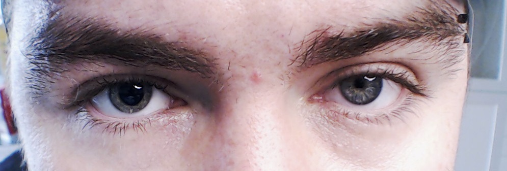
- Pupil sizes measured in both bright light and dim light – this comparison is the core of the examination
- Anisocoria greater in the dark indicates the smaller pupil is abnormal (sympathetic defect)
- Anisocoria greater in the light indicates the larger pupil is abnormal (parasympathetic defect)
- Equal difference in light and dark, with brisk reactions, indicates physiological anisocoria
- Ptosis – mild with Horner syndrome, often marked with a third nerve palsy
- Eye movements and lid position to identify a third nerve palsy
- Slit-lamp assessment for iris trauma, synechiae, or the segmental vermiform iris movements of an Adie pupil
Measuring the pupils in the dark is the step most often skipped and the one that decides the entire diagnostic direction.
Diagnostic Evaluation
Pharmacological Testing
- Low-concentration pilocarpine constricts an Adie tonic pupil through denervation supersensitivity, but not a normal pupil
- Full-strength pilocarpine constricts a pupil with a third nerve palsy but fails to constrict a pharmacologically blocked pupil
- Apraclonidine reverses the anisocoria of Horner syndrome, and has largely replaced cocaine testing in practice
Urgent Imaging
- A pupil-involving third nerve palsy requires urgent CT or MR angiography to exclude a posterior communicating artery aneurysm
- Horner syndrome requires imaging of the entire oculosympathetic pathway – brain, neck with carotid vessels, and lung apex
- Painful Horner syndrome should be treated as a possible carotid dissection and imaged emergently
Paediatric Considerations
- Horner syndrome in a child requires evaluation for neuroblastoma, including imaging of the neck, chest and abdomen and urinary catecholamine testing
- Heterochromia with a lighter iris on the affected side suggests a congenital sympathetic lesion
The rule that governs urgency is simple: a new pupil-involving third nerve palsy and a painful Horner syndrome are both same-day problems.
Differential Diagnosis
The differential is really a structured decision about which pupil is abnormal:
- Physiological anisocoria – equal difference in light and dark, normal lids and movements
- Horner syndrome – miosis with mild ptosis, worse in dark, apraclonidine-positive
- Third nerve palsy – mydriasis with ptosis and an eye deviated down and out
- Adie tonic pupil – large pupil, poor light reaction, slow tonic near response, supersensitive to dilute pilocarpine
- Pharmacological mydriasis – a very large pupil that will not constrict even to full-strength pilocarpine
- Iris pathology – synechiae, trauma or previous surgery visible at the slit lamp
Light-near dissociation – a pupil that reacts far better to near than to light – narrows the field quickly to Adie pupil, Argyll Robertson pupils and dorsal midbrain pathology.
Management
Emergency Pathways
- Pupil-involving third nerve palsy: immediate neuroimaging with angiography and urgent neurosurgical involvement
- Painful or acute Horner syndrome: emergency imaging for carotid dissection, with stroke-prevention treatment as indicated
- Acute angle-closure glaucoma: immediate pressure-lowering treatment and laser iridotomy
Non-Urgent Management
- Physiological anisocoria requires only reassurance and documentation
- Adie tonic pupil is managed symptomatically, with reading correction and tinted lenses where glare is troublesome; dilute pilocarpine can be used cosmetically
- Pharmacological anisocoria resolves once the agent is identified and withdrawn
- Iritis is treated with topical steroid and cycloplegia to break synechiae
Documentation
- Record pupil sizes in light and dark in millimetres rather than describing them qualitatively
- Photograph the pupils where possible, since a baseline image is invaluable if the patient re-presents
Most anisocoria needs no treatment at all – but that conclusion is only safe once the dangerous causes have been actively excluded rather than assumed away.
Prognosis
Outcome is entirely determined by the underlying cause.
- Physiological anisocoria is stable lifelong and carries no consequence
- Adie tonic pupil is benign; the pupil often becomes smaller over years, and accommodation improves
- Pharmacological anisocoria resolves fully once the agent clears
- A compressive third nerve palsy from aneurysm carries a serious prognosis that depends entirely on how quickly it is diagnosed and treated
- Horner syndrome itself is harmless; its prognosis is that of the lesion causing it, which may be a dissection or a malignancy
Anisocoria is best thought of as a question rather than a finding – benign in most patients, but occasionally the earliest visible sign of something that will not wait.


Document what you see
Two smartphone imaging tools built for everyday clinic use — one for the slit lamp, one for the fundus.
From Choroida — the team behind this siteReferences
- Kardon R, Thompson HS. The pupil. In: Walsh and Hoyt’s Clinical Neuro-Ophthalmology.
- Bremner F. Pupil assessment in optic nerve disorders. Eye. 2004.
- Trobe JD. The evaluation of anisocoria. Journal of Neuro-Ophthalmology.
- Anisocoria. StatPearls, NCBI Bookshelf.
- Anisocoria. EyeWiki, American Academy of Ophthalmology.
Test yourself
A few questions straight from this article.
-
Anisocoria that is greater in the dark localises the problem to which pupil and pathway?
Anisocoria greater in the dark means the smaller pupil is failing to dilate, indicating a sympathetic defect such as Horner syndrome; a difference greater in light implicates the larger pupil instead. -
How common is physiological anisocoria in the normal population?
Physiological anisocoria is present in roughly a fifth of the normal population and is the single commonest cause, showing an equal difference in light and dark with normal reactions. -
Why does a compressive third nerve lesion involve the pupil early while an ischaemic one spares it?
The parasympathetic fibres travel on the outer surface of the third cranial nerve, so an external compressive lesion affects the pupil early, whereas ischaemic microvascular damage typically spares it. -
What does a new pupil-involving third nerve palsy require?
A pupil-involving third nerve palsy is a posterior communicating artery aneurysm until proven otherwise, requiring immediate angiography and urgent neurosurgical involvement. -
What should a painful Horner syndrome be treated as until proven otherwise?
Painful Horner syndrome should be treated as a possible carotid dissection and imaged emergently, with stroke-prevention treatment as indicated. -
Which pharmacological response confirms an Adie tonic pupil?
Low-concentration pilocarpine constricts an Adie tonic pupil through denervation supersensitivity of the ciliary ganglion, while leaving a normal pupil unchanged. -
Which agent reverses the anisocoria of Horner syndrome in modern practice?
Apraclonidine reverses the anisocoria of Horner syndrome and has largely replaced cocaine testing in practice. -
Which condition must be excluded when Horner syndrome is found in a child?
Horner syndrome in a child requires evaluation for neuroblastoma, with imaging of the neck, chest and abdomen plus urinary catecholamine testing. -
Light-near dissociation in an unequal pupil narrows the differential to which group?
A pupil that reacts far better to near than to light narrows the field quickly to Adie pupil, Argyll Robertson pupils and dorsal midbrain pathology. -
What determines the prognosis of a patient found to have Horner syndrome?
Horner syndrome is harmless in itself; its prognosis is entirely that of the underlying lesion, which may be a carotid dissection or a malignancy.

Writes and reviews the clinical articles on Educate. Every post is checked against peer-reviewed sources before it goes live.


