
An episcleral hemangioma is a vascular proliferation that can occur as an isolated finding, but its association with Sturge-Weber syndrome is significant enough that identifying one should prompt a careful look for the broader facial and systemic features of that syndrome, particularly since the glaucoma risk in Sturge-Weber syndrome is substantial and benefits from early recognition and surveillance.
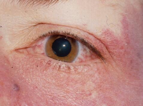
What an Episcleral Hemangioma Is
This is a benign vascular tumor arising within the episclera, the vascularized connective tissue layer between the conjunctiva and the sclera, appearing as a reddish, vascular-appearing mass or area of diffuse vascular proliferation on the surface of the eye.
The Sturge-Weber Syndrome Association
Sturge-Weber syndrome is a neurocutaneous syndrome characterized by a facial capillary malformation (port-wine stain), typically in the distribution of the trigeminal nerve, along with leptomeningeal vascular malformation and, in the eye, a spectrum of vascular findings that can include episcleral hemangioma, choroidal hemangioma, and glaucoma.
- When an episcleral hemangioma is identified, particularly in a patient with a facial port-wine stain or other suggestive features, evaluation for the broader Sturge-Weber syndrome spectrum, including choroidal hemangioma (see phakomatoses ocular manifestations for the broader context of this and related neurocutaneous syndromes) and glaucoma, is warranted
- Isolated episcleral hemangioma without other features of Sturge-Weber syndrome also occurs, representing a localized vascular anomaly without the broader systemic and intracranial associations
Fundus Explorer Pro
Photograph the retinal findings described here with the phone already in your pocket — 22 D optics and built-in illumination in one handheld unit.
From Choroida — the team behind this siteGlaucoma Risk
Glaucoma is a common and clinically significant complication in Sturge-Weber syndrome, related in part to elevated episcleral venous pressure from the associated vascular malformations affecting normal aqueous outflow dynamics (see elevated episcleral venous pressure glaucoma for a detailed discussion of this specific mechanism).
- Glaucoma associated with Sturge-Weber syndrome can present either in infancy, sometimes with buphthalmos similar to primary congenital glaucoma, or later in childhood or adulthood, with a more gradual onset
- Given this significant, well-documented glaucoma risk, any patient identified with an episcleral hemangioma, particularly in the context of broader Sturge-Weber syndrome features, warrants prompt and then ongoing intraocular pressure surveillance
Clinical Presentation
- A visible, reddish, vascular-appearing mass or diffuse area of prominent, dilated episcleral vessels on the surface of the eye
- Associated dilated, tortuous episcleral vessels more broadly, reflecting the elevated episcleral venous pressure often present in the broader syndrome
- Facial port-wine stain, when present, typically ipsilateral to the affected eye, supporting the Sturge-Weber syndrome association
- Signs of elevated intraocular pressure or, in an infant, buphthalmos, when glaucoma has already developed
Diagnostic Evaluation
- Careful external and slit-lamp examination characterizing the episcleral vascular finding and assessing for dilated, tortuous episcleral vessels more broadly
- A thorough assessment for facial port-wine stain and other features suggestive of Sturge-Weber syndrome
- Dilated fundus examination to assess for an associated choroidal hemangioma, another recognized ocular manifestation of this syndrome
- Intraocular pressure measurement, with particular vigilance and appropriately frequent monitoring given the significant associated glaucoma risk
- Neuroimaging and broader systemic evaluation when Sturge-Weber syndrome is suspected, given the potential for associated intracranial and other systemic involvement
Management
Managing the Episcleral Hemangioma Itself
The episcleral hemangioma itself generally does not require direct treatment, since it is a benign vascular lesion, though very rarely, treatment may be considered for significant cosmetic concern or if it is contributing meaningfully to symptomatic ocular surface irritation.
Managing Associated Glaucoma
Glaucoma associated with Sturge-Weber syndrome can be challenging to manage given the underlying elevated episcleral venous pressure mechanism, generally beginning with medical aqueous-suppressant therapy, with surgical intervention considered for inadequately controlled disease, and with specific attention to the increased surgical risks (including choroidal effusion) that elevated episcleral venous pressure conditions carry for intraocular surgery.
Association with Sturge-Weber syndrome
An episcleral hemangioma is often associated with the Sturge-Weber syndrome, with a port-wine stain on the face, glaucoma, and a diffuse choroidal hemangioma. A child with an episcleral lesion needs careful assessment of intraocular pressure, the optic disc, and the fundus (see Sturge-Weber syndrome). Elevated episcleral venous pressure is a cause of glaucoma in these eyes.
Follow-up
Check the pressure at regular intervals, and examine the fundus under dilation. Imaging with ultrasound or OCT helps in assessing choroidal involvement. Reassure parents that the lesion is benign, and explain that the pressure is the important point.
Prognosis
The episcleral hemangioma itself carries an excellent prognosis, being a benign vascular finding without direct threat to vision on its own.
The broader prognosis, when associated with Sturge-Weber syndrome, depends substantially on how effectively the associated glaucoma is monitored and managed over a patient’s lifetime, underscoring why identifying this specific vascular finding, and pursuing the systemic evaluation it should prompt, carries real, lasting clinical importance beyond the finding itself.


Document what you see
Two smartphone imaging tools built for everyday clinic use — one for the slit lamp, one for the fundus.
From Choroida — the team behind this siteReferences
- Sullivan TJ, Clarke MP, Morin JD. The ocular manifestations of the Sturge-Weber syndrome. J Pediatr Ophthalmol Strabismus. 1992;29:349-356.
- Sujansky E, Conradi S. Sturge-Weber syndrome: age of onset of seizures and glaucoma and the prognosis for affected children. J Child Neurol. 1995;10:49-58.
- Mantelli F, Bruscolini A, La Cava M, Abdolrahimzadeh S, Lambiase A. Ocular manifestations of Sturge-Weber syndrome: pathogenesis, diagnosis, and management. Clin Ophthalmol. 2016;10:871-878.
- Phelps CD. The pathogenesis of glaucoma in Sturge-Weber syndrome. Ophthalmology. 1978;85:276-286.
