
Disciform keratitis marks a genuine turning point in herpetic eye disease, since it represents the immune system’s response to viral antigen rather than active viral replication itself, and this distinction is precisely why its treatment, built around corticosteroids rather than antivirals alone, differs so fundamentally from the management of the epithelial dendrite that may have preceded it.
A Different Disease Process From Epithelial Herpetic Keratitis
Epithelial herpes simplex keratitis, with its characteristic dendritic ulcer, represents active viral replication within the corneal epithelium, and treatment is centered on antiviral therapy to halt this replication.
Disciform keratitis, by contrast, reflects an immune-mediated inflammatory response, most often centered on the corneal endothelium, occurring in response to herpetic antigen, sometimes following a prior episode of epithelial disease but sometimes occurring without any preceding epithelial involvement, which is why understanding this as an immunologic rather than directly infectious process is central to managing it correctly.
Clinical Features
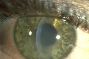
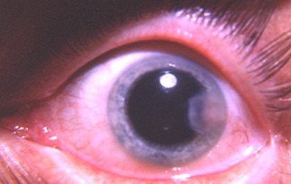
- A characteristic disc-shaped (hence “disciform”) area of stromal edema, usually central or paracentral, giving the cornea a localized, rounded area of haze and swelling
- Keratic precipitates, often concentrated directly beneath the area of stromal edema, reflecting the endothelial involvement that is central to this condition’s mechanism
- Reduced vision correlating with the degree of stromal and epithelial edema
- Mild to moderate discomfort and photophobia, generally less severe than the pain typical of active epithelial disease
- A history of prior herpetic eye disease is common, though disciform keratitis can occur as an initial presentation in some patients
- Elevated intraocular pressure can accompany disciform keratitis, related to associated trabeculitis, and should be specifically assessed given the implications for concurrent glaucoma management
All-fit Slit-Lamp Adapter
Record and share exactly what you see at the slit lamp. One adapter fits any slit lamp or surgical microscope — and any smartphone.
From Choroida — the team behind this siteDistinguishing From Active Epithelial Disease
Careful slit-lamp examination, including fluorescein staining, is important to determine whether any active epithelial dendritic or geographic ulceration is present concurrently, since this distinction directly shapes the treatment approach: active epithelial disease requires antiviral treatment as a primary measure, while isolated disciform (stromal/endothelial) disease without active epithelial ulceration is managed primarily as an immune-mediated process.
Diagnostic Evaluation
- Slit-lamp examination characterizing the pattern and depth of stromal edema, the distribution of keratic precipitates, and careful assessment for any concurrent active epithelial disease
- Intraocular pressure measurement, given the recognized association with trabeculitis and secondary pressure elevation
- A history of prior herpetic ocular disease episodes, though the absence of such a history does not exclude the diagnosis
Management
Topical Corticosteroids
Topical corticosteroids are the central treatment for disciform keratitis, addressing the underlying immune-mediated inflammation directly, typically started at a moderate dose and gradually tapered over weeks based on clinical response, since inflammation can rebound if steroids are withdrawn too quickly.
Antiviral Cover
Even though disciform keratitis itself is immune-mediated rather than directly infectious, concurrent antiviral therapy is generally used alongside corticosteroids, both to treat any coexisting active epithelial disease and as prophylaxis against corticosteroid-associated reactivation of viral replication, since topical corticosteroids used without antiviral cover in a herpetic eye carry a recognized risk of precipitating a more severe epithelial flare.
Managing Associated Elevated Intraocular Pressure
When trabeculitis has caused secondary pressure elevation, aqueous-suppressant glaucoma therapy is added as needed, with treatment of the underlying inflammatory disciform process itself remaining central to resolving this associated pressure elevation over time.
Long-Term Suppressive Therapy
Given the recurrent nature of herpetic eye disease, long-term oral antiviral suppressive therapy is often considered, particularly after a significant or recurrent episode of disciform keratitis, aiming to reduce the frequency of future recurrences and their cumulative impact on corneal clarity and endothelial health.
Why Recurrence Matters
Each episode of disciform keratitis carries some risk of cumulative endothelial cell loss and stromal scarring, and recurrent episodes over a patient’s lifetime can progressively threaten corneal clarity, which is the main long-term driver of vision loss in chronic herpetic eye disease and the central reason both prompt treatment of active episodes and consideration of suppressive therapy matter for long-term visual preservation.
Recurrence prevention
The Herpetic Eye Disease Study showed that long-term oral acyclovir reduces recurrences of herpetic eye disease. Many patients with disciform keratitis benefit from prophylaxis, particularly after repeated episodes, and the dose and duration are chosen with kidney function in mind. Topical steroids are always used with antiviral cover, and tapered slowly.
Prognosis
Individual episodes of disciform keratitis generally respond well to appropriately balanced corticosteroid and antiviral treatment, with most patients experiencing significant improvement in stromal edema and vision as inflammation resolves.
The greater long-term concern is the cumulative effect of recurrent episodes on corneal clarity and endothelial function, underscoring the value of antiviral suppressive therapy in patients with a pattern of recurrent disease, and ongoing surveillance for both recurrence and the chronic sequelae of repeated inflammatory episodes.


Document what you see
Two smartphone imaging tools built for everyday clinic use — one for the slit lamp, one for the fundus.
From Choroida — the team behind this siteReferences
- Liesegang TJ. Herpes simplex virus epidemiology and ocular importance. Cornea. 2001;20:1-13.
- Wilhelmus KR, Gee L, Hauck WW, et al. Herpetic Eye Disease Study. A controlled trial of topical corticosteroids for herpes simplex stromal keratitis. Ophthalmology. 1994;101:1883-1895.
- Herpetic Eye Disease Study Group. Acyclovir for the prevention of recurrent herpes simplex virus eye disease. N Engl J Med. 1998;339:300-306.
- Holland EJ, Schwartz GS. Classification of herpes simplex virus keratitis. Cornea. 1999;18:144-154.
