
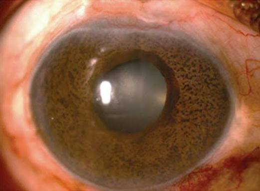
Cogan-Reese syndrome produces a finding that can look genuinely alarming at first glance, multiple small, pigmented, sometimes pedunculated nodules studding the iris surface, and correctly recognizing these as a manifestation of the broader iridocorneal endothelial syndrome spectrum, rather than a primary melanocytic iris tumor, is essential given how differently these two possibilities are managed.
Where Cogan-Reese Fits in the ICE Spectrum
Cogan-Reese syndrome, sometimes called iris nevus syndrome, is the third recognized presentation within the iridocorneal endothelial syndrome spectrum, alongside essential iris atrophy and Chandler syndrome, sharing the same underlying mechanism: an abnormal corneal endothelial cell population with epithelial-like behavior that proliferates across the angle and onto the iris surface.
In Cogan-Reese syndrome specifically, this abnormal membrane growing across the iris surface entraps and distorts normal iris stromal tissue as it contracts, producing the characteristic pigmented nodules that give this presentation its name.
What the Nodules Actually Are
The pigmented nodules of Cogan-Reese syndrome are not a primary neoplastic proliferation of melanocytes, as a true iris nevus or melanoma would be, but rather represent entrapped and distorted iris stromal tissue, pulled up into nodular projections by the contracting abnormal endothelial membrane spreading across the iris surface, a fundamentally mechanical rather than neoplastic process.
All-fit Slit-Lamp Adapter
Record and share exactly what you see at the slit lamp. One adapter fits any slit lamp or surgical microscope — and any smartphone.
From Choroida — the team behind this siteClinical Features
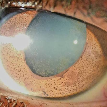
- Multiple small, pigmented nodules on the iris surface, which can appear pedunculated (having a stalk-like base) or sessile, and vary in size and number between affected patients
- Progressive corectopia and iris stromal atrophy can accompany the nodules, reflecting the same contractile membrane process that produces these changes in the other ICE syndrome variants, though typically less severe than the dramatic changes of essential iris atrophy
- Corneal edema can occur, related to the same underlying abnormal endothelium, similarly to Chandler syndrome
- Peripheral anterior synechiae and secondary glaucoma are common, given the angle involvement shared across the ICE syndrome spectrum
- Almost always unilateral, consistent with the broader pattern seen throughout this syndrome spectrum
Distinguishing From Iris Melanoma and Other Pigmented Lesions
This distinction carries genuine clinical weight, since the management implications of a benign ICE syndrome manifestation versus a primary melanocytic iris tumor are entirely different.
- Multiple small nodules, rather than a single dominant, growing pigmented mass, favor Cogan-Reese syndrome over a primary melanocytic tumor
- Accompanying findings characteristic of the broader ICE syndrome, including corneal edema, an abnormal endothelial appearance on specular or confocal microscopy, and peripheral anterior synechiae on gonioscopy, support the diagnosis of Cogan-Reese syndrome rather than an isolated iris tumor
- Unlike a true iris melanoma, the nodules of Cogan-Reese syndrome are generally stable or only slowly changing in the context of the broader, gradually evolving ICE syndrome process, rather than showing the more concerning growth pattern that would prompt urgent oncologic evaluation of a suspected melanoma (see iris nevus versus melanoma for the general risk-feature framework used to evaluate a concerning pigmented iris lesion)
Diagnostic Evaluation
- Slit-lamp examination characterizing the nodules alongside assessment of corneal clarity and any iris stromal atrophy or corectopia
- Specular or confocal microscopy to identify the characteristic abnormal endothelial cell pattern shared across the ICE syndrome spectrum
- Gonioscopy to assess angle involvement and peripheral anterior synechiae
- Intraocular pressure monitoring, given the meaningful association with secondary glaucoma
Management
Managing Secondary Glaucoma
As with the other ICE syndrome variants, glaucoma management is central to care when present, beginning with aqueous-suppressant medical therapy and progressing to surgical intervention, generally favoring glaucoma drainage device implantation over trabeculectomy, for inadequately controlled disease.
Managing Corneal Edema
Hypertonic saline supports mild to moderate edema symptomatically, while endothelial keratoplasty is considered for more significant, visually limiting corneal decompensation, with the same caveat about potential ongoing abnormal endothelial activity affecting graft longevity that applies across the ICE syndrome spectrum.
Ongoing Monitoring
Regular follow-up tracking the nodules, corneal clarity, and intraocular pressure supports early identification of any disease progression requiring treatment adjustment over time.
A quick clinical pearl
Look at the iris with a narrow beam in each quadrant. The pedunculated, dark nodules can be sparse and subtle, and the pupil may be displaced toward the area of greatest atrophy. Photographs help to monitor change.
Prognosis
Prognosis in Cogan-Reese syndrome relates primarily to the degree of secondary glaucoma and corneal involvement present, following a broadly similar overall trajectory to the other ICE syndrome variants, chronic and slowly progressive, but generally manageable with sustained, vigilant care.
Correctly recognizing the pigmented nodules as part of this syndrome, rather than a primary melanocytic tumor requiring oncologic workup, avoids unnecessary anxiety and intervention while still supporting the ongoing glaucoma and corneal surveillance this chronic condition genuinely requires.


Document what you see
Two smartphone imaging tools built for everyday clinic use — one for the slit lamp, one for the fundus.
From Choroida — the team behind this siteReferences
- Shields MB. Progressive essential iris atrophy, Chandler’s syndrome, and the iris nevus (Cogan-Reese) syndrome: a spectrum of disease. Surv Ophthalmol. 1979;24:3-20.
- Cogan DG, Reese AB. A syndrome of iris nodules, ectopic Descemet’s membrane, and unilateral glaucoma. Doc Ophthalmol. 1969;26:424-433.
- Alvarado JA, Murphy CG, Maglio M, Hetherington J. Pathogenesis of Chandler’s syndrome, essential iris atrophy and the Cogan-Reese syndrome. Invest Ophthalmol Vis Sci. 1986;27:853-872.
- Shields JA, Shields CL. Atlas of Intraocular Tumors. 2nd ed. Philadelphia: Lippincott Williams & Wilkins; 2008.
Test yourself
A few questions straight from this article.
-
What are the pigmented iris nodules of Cogan-Reese syndrome made of?
The abnormal endothelial membrane spreading across the iris entraps and distorts normal iris stroma as it contracts, pulling it into nodular projections: a mechanical rather than neoplastic process. -
Which nodule pattern favours Cogan-Reese syndrome over a primary melanocytic iris tumour?
Multiple small nodules studding the iris favour Cogan-Reese syndrome, whereas a single dominant, growing pigmented mass points towards a primary melanocytic tumour. -
How do the iris nodules of Cogan-Reese syndrome typically behave over time?
The nodules are generally stable or only slowly changing within the gradually evolving ICE process, unlike the growth pattern that would prompt urgent oncologic evaluation of a suspected melanoma. -
Which description matches the iris nodules seen in Cogan-Reese syndrome?
The nodules are multiple, small and pigmented, may be pedunculated with a stalk-like base or sessile, and vary in size and number between affected patients. -
Which complications are common in Cogan-Reese syndrome?
Peripheral anterior synechiae and secondary glaucoma are common because of the angle involvement shared across the iridocorneal endothelial syndrome spectrum. -
Which glaucoma operation is generally favoured in Cogan-Reese syndrome when medical therapy fails?
Management starts with aqueous-suppressant medical therapy and progresses to surgery for inadequately controlled disease, generally favouring a glaucoma drainage device over trabeculectomy. -
Which additional iris changes can accompany the nodules in Cogan-Reese syndrome?
The same contractile membrane process can produce progressive corectopia and iris stromal atrophy, though these are typically less severe than the dramatic changes of essential iris atrophy. -
Which examination approach best detects the sparse iris nodules of Cogan-Reese syndrome?
The dark, pedunculated nodules can be sparse and subtle, so the iris should be examined with a narrow beam in each quadrant; photographs help monitor change. -
Cogan-Reese syndrome is also known by which other name?
Cogan-Reese syndrome is sometimes called iris nevus syndrome, although the nodules are entrapped iris stroma rather than a true melanocytic nevus. -
Prognosis in Cogan-Reese syndrome depends primarily on which factors?
Prognosis relates primarily to how much secondary glaucoma and corneal involvement are present; the course is chronic and slowly progressive but generally manageable with vigilant care.

Writes and reviews the clinical articles on Educate. Every post is checked against peer-reviewed sources before it goes live.


