
Cyclophotocoagulation lowers intraocular pressure by directly ablating the ciliary body’s aqueous-producing epithelium with laser energy, reducing the amount of fluid the eye produces rather than working to improve outflow the way trabeculectomy, tube shunt surgery, and MIGS procedures, all discussed in their own dedicated articles on this site, do.
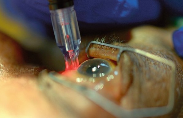
That fundamental difference in mechanism, reducing production rather than improving drainage, is what has historically defined cyclophotocoagulation’s role as a treatment reserved for eyes with limited remaining options, though newer, gentler versions of the technique have begun to expand that role somewhat.
Understanding the different delivery techniques, and why some are considered more destructive than others, explains both the procedure’s traditional reputation and its evolving place in modern glaucoma management.
The ciliary body’s dual role, producing aqueous while also supporting other ocular functions, is part of why deliberately damaging it carries a genuinely different risk calculus than the outflow-focused procedures more commonly used earlier in the glaucoma treatment ladder.
Mechanism
The ciliary body’s non-pigmented epithelium is responsible for aqueous humor production, and laser energy delivered to this tissue causes coagulative damage that reduces its secretory capacity, directly lowering the rate of aqueous inflow into the eye.
Because this approach reduces production rather than addressing outflow, it works regardless of the underlying cause of a given eye’s elevated pressure, which is part of why it has traditionally been reserved for refractory glaucoma where outflow-based procedures have already failed or are not expected to succeed.
The degree of pressure reduction depends on how much ciliary body tissue is effectively treated, which is influenced by the specific technique, energy settings, and treatment extent chosen for a given eye.
Because the effect is not immediately titratable the way a topical medication dose can be adjusted day to day, some patients need more than one treatment session over time to reach and maintain their target pressure.
Delivery Techniques
Transscleral cyclophotocoagulation delivers laser energy through the sclera from outside the eye, without direct visualization of the ciliary body itself, using anatomic landmarks to guide probe placement over the pars plicata.
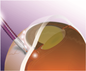
Endoscopic cyclophotocoagulation uses a small camera and laser probe inserted into the eye, allowing the surgeon to directly visualize and treat the ciliary processes under controlled conditions, often combined with cataract surgery in the same session.
Micropulse transscleral cyclophotocoagulation delivers the same transscleral energy in short, repeated pulses with rest periods between them, aiming to reduce collateral thermal damage to adjacent tissue compared to continuous-wave delivery while still achieving a meaningful pressure-lowering effect.
All-fit Slit-Lamp Adapter
Record and share exactly what you see at the slit lamp. One adapter fits any slit lamp or surgical microscope — and any smartphone.
From Choroida — the team behind this siteTraditional Versus Modern Role
- Continuous-wave transscleral cyclophotocoagulation has traditionally been reserved for eyes with refractory glaucoma and limited visual potential, given its more destructive nature and less predictable, sometimes more severe complication profile
- Endoscopic cyclophotocoagulation is used more often in eyes with reasonable visual potential, particularly when combined with cataract surgery, given its more controlled, directly visualized approach
- Micropulse cyclophotocoagulation has expanded the technique’s use into eyes with better visual potential and even earlier-stage glaucoma, given its gentler energy delivery and generally more favorable safety profile compared to continuous-wave treatment
Complications
Inflammation and pain are common in the immediate postoperative period, generally managed with topical corticosteroids and appropriate analgesia.
Hypotony, sometimes significant, is a recognized complication, particularly with more aggressive continuous-wave treatment, reflecting the risk of overtreating the ciliary body’s secretory capacity relative to what a given eye actually needs.
Vision loss, while less common with the gentler modern techniques than with traditional continuous-wave treatment, remains a real risk that needs to be discussed clearly with patients before proceeding, particularly for eyes with reasonable baseline visual potential.
Patient Selection
Traditional cyclophotocoagulation candidates include eyes with advanced, refractory glaucoma that has failed multiple prior surgical interventions, or eyes with limited visual potential where a more aggressive, less predictable procedure carries an acceptable risk-benefit balance.
Newer micropulse and endoscopic techniques have broadened candidacy to include eyes earlier in their glaucoma course, or those undergoing cataract surgery who could benefit from an additional pressure-lowering procedure performed in the same setting.
The choice of technique, and whether cyclophotocoagulation is appropriate at all for a given eye, depends on the stage of glaucoma, prior treatment history, visual potential, and the specific pressure target needed, following the same individualized decision-making used across the broader spectrum of glaucoma surgical options.
Patients considering the procedure benefit from a clear, honest explanation of which specific technique is being proposed, since the historical reputation of “cyclophotocoagulation” as a last-resort, high-risk treatment applies far more to older continuous-wave transscleral delivery than to the gentler modern variants now in wider use.


Document what you see
Two smartphone imaging tools built for everyday clinic use — one for the slit lamp, one for the fundus.
From Choroida — the team behind this siteReferences
- American Academy of Ophthalmology. Basic and Clinical Science Course, Section 10: Glaucoma.
- Aquino MC, Barton K, Tan AM, et al. Micropulse versus continuous wave transscleral diode cycloablation study: a randomised clinical trial. Clinical and Experimental Ophthalmology.
- Francis BA, Kawji AS, Vo NT, et al. Endoscopic cyclophotocoagulation (ECP) in the management of uncontrolled glaucoma with prior aqueous tube shunt. Journal of Glaucoma.
Test yourself
A few questions straight from this article.
-
Which glaucoma procedure lowers pressure by reducing aqueous production rather than improving outflow?
Cyclophotocoagulation ablates the ciliary body's aqueous-producing epithelium, reducing how much fluid the eye makes, while trabeculectomy, tube shunts and MIGS all work by improving outflow. -
Which tissue does cyclophotocoagulation ablate to lower intraocular pressure?
The non-pigmented ciliary epithelium produces aqueous humor, and laser energy delivered to it causes coagulative damage that reduces its secretory capacity and lowers aqueous inflow. -
Why do some patients need more than one cyclophotocoagulation session?
The effect is not immediately titratable the way a topical medication dose can be adjusted day to day, so some patients need more than one session over time to reach and maintain their target pressure. -
What characterizes transscleral cyclophotocoagulation?
Transscleral delivery sends laser energy through the sclera from outside the eye, without direct visualization of the ciliary body, using anatomic landmarks to guide probe placement over the pars plicata. -
What is the main advantage of endoscopic cyclophotocoagulation?
A small camera and laser probe are inserted into the eye so the surgeon can directly visualize and treat the ciliary processes under controlled conditions, often combined with cataract surgery in the same session. -
How does micropulse transscleral delivery differ from continuous-wave delivery?
Micropulse delivers the same transscleral energy in short repeated pulses with rest periods between them, aiming to reduce collateral thermal damage to adjacent tissue while still lowering pressure meaningfully. -
Which technique has traditionally been reserved for refractory glaucoma with limited visual potential?
Continuous-wave transscleral treatment has traditionally been reserved for refractory glaucoma with limited visual potential, given its more destructive nature and less predictable, sometimes more severe complication profile. -
Which eyes became candidates as gentler cyclophotocoagulation techniques emerged?
Micropulse and endoscopic techniques have broadened candidacy to eyes earlier in their glaucoma course, and to eyes undergoing cataract surgery that could benefit from an added pressure-lowering procedure in the same setting. -
Which complication reflects overtreatment of the ciliary body's secretory capacity?
Hypotony is a recognized complication, particularly with more aggressive continuous-wave treatment, reflecting the risk of overtreating the ciliary body's secretory capacity relative to what the eye actually needs. -
What should patients be told about cyclophotocoagulation's last-resort reputation?
The historical reputation as a last-resort, high-risk treatment applies far more to older continuous-wave transscleral delivery than to the gentler modern variants, so patients benefit from knowing which technique is being proposed.

Writes and reviews the clinical articles on Educate. Every post is checked against peer-reviewed sources before it goes live.


