
Coats-like retinal telangiectasia describes a pattern of abnormal, dilated, and often saccular retinal blood vessels that leak lipid-rich fluid into and beneath the retina, and recognizing this specific vascular pattern matters urgently in a young child, since the resulting exudation and retinal detachment can produce leukocoria, a white pupillary reflex that must always be distinguished promptly from the far more serious diagnosis it can mimic, retinoblastoma.
What’s Wrong With the Vessels
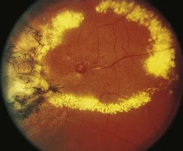
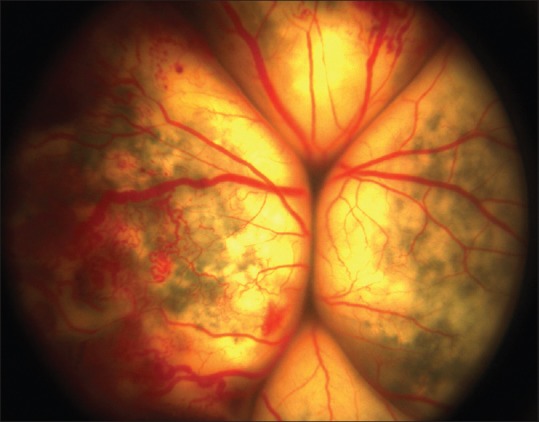
The retinal telangiectasia at the heart of this condition consists of abnormally dilated, tortuous retinal vessels with saccular or fusiform outpouchings, classically described as having a “light bulb” appearance on careful examination, and these abnormal vessels have compromised walls that allow lipid-rich plasma to leak into the surrounding retina and subretinal space.
Coats Disease
Coats disease is the classic, most extensively described condition producing this pattern, typically presenting in childhood, predominantly in males, and almost always unilateral, a notable and useful distinguishing feature from many other pediatric retinal conditions that tend toward bilateral involvement.
- Progressive telangiectasia and exudation, accumulating as yellow, often extensive subretinal and intraretinal lipid deposits, sometimes forming a macular lipid star
- Advanced disease can produce massive subretinal exudation and exudative retinal detachment, which is what produces the leukocoria that often brings the child to medical attention
- The absence of a discrete mass distinguishes Coats disease from retinoblastoma on careful examination and imaging, even though both can present with a strikingly similar white pupillary reflex to a casual observer
Fundus Explorer Pro
Photograph the retinal findings described here with the phone already in your pocket — 22 D optics and built-in illumination in one handheld unit.
From Choroida — the team behind this siteThe Critical Differential: Retinoblastoma
Any child presenting with leukocoria requires urgent, careful evaluation to distinguish Coats disease from retinoblastoma, since the two conditions require entirely different management and retinoblastoma carries life-threatening implications if missed or delayed.
- Retinoblastoma typically shows a discrete retinal mass on examination and imaging, often with calcification detectable on ultrasound or CT, features not present in Coats disease
- Coats disease shows the characteristic telangiectatic vessels and lipid exudation pattern without a discrete solid tumor mass
- When the distinction is not immediately clear on clinical examination alone, ultrasound, and in some cases MRI, along with examination under anesthesia, are used to confirm the diagnosis before proceeding with any treatment, given how consequential distinguishing these two conditions correctly is
Clinical Presentation
- Leukocoria or strabismus are the most common presenting signs, particularly in more advanced disease with significant exudation or detachment
- Younger children may be brought in specifically because a parent or caregiver, or a photograph, noticed an abnormal white reflex in one eye
- Older children and, less commonly, adults can present with a milder, incidentally discovered form of retinal telangiectasia and exudation, sometimes discovered on routine examination before advancing to the more severe presentation typical of classic pediatric Coats disease
Diagnostic Evaluation
- Dilated fundus examination, looking specifically for the characteristic telangiectatic “light bulb” vessels and pattern of lipid exudation
- Fluorescein angiography is valuable for clearly delineating the abnormal telangiectatic vessels and areas of leakage, helping to guide treatment planning
- Ultrasound to assess for any retinal detachment, characterize the extent of exudation, and importantly, to exclude calcification that would instead suggest retinoblastoma
- Examination under anesthesia is often necessary in young children to obtain an adequate, complete examination and to correctly stage the disease
Management
Ablative Treatment of Abnormal Vessels
Laser photocoagulation or cryotherapy directed at the telangiectatic vessels is the primary treatment approach, aiming to close off the abnormal, leaking vessels and halt further exudation, often requiring more than one treatment session depending on disease extent.
Managing Exudation and Detachment
In eyes with significant subretinal fluid or exudative detachment, treatment of the causative vessels is generally still the primary approach, since successfully closing the abnormal vessels often allows gradual resorption of existing subretinal fluid and exudate over subsequent months, though very advanced, total detachment may have a more limited visual prognosis despite treatment.
Staging and Prognosis-Based Decisions
Disease staging based on the extent of telangiectasia, exudation, and detachment informs both prognosis and the intensity of treatment pursued, with earlier-stage disease generally carrying a substantially better visual outcome than advanced, total exudative detachment.
Prognosis
Outcomes in Coats disease depend heavily on the stage at diagnosis, with earlier detection and treatment, before extensive exudation and detachment develop, offering the best chance of preserving useful vision in the affected eye.
Even in advanced disease where visual prognosis is limited, appropriate treatment remains valuable for globe preservation and comfort, and correctly distinguishing the condition from retinoblastoma at the outset remains the single most consequential diagnostic step in the entire evaluation.


Document what you see
Two smartphone imaging tools built for everyday clinic use — one for the slit lamp, one for the fundus.
From Choroida — the team behind this siteReferences
- Shields JA, Shields CL. Review: Coats disease: the 2001 LuEsther T. Mertz lecture. Retina. 2002;22:80-91.
- Shields JA, Shields CL, Honavar SG, Demirci H. Clinical variations and complications of Coats disease in 150 cases: the 2000 Sanford Gifford Memorial Lecture. Am J Ophthalmol. 2001;131:561-571.
- Ghorbanian S, Jaulim A, Chatziralli IP. Diagnosis and treatment of Coats’ disease: a review of the literature. Ophthalmologica. 2012;227:175-182.
- Shields CL, Uysal Y, Benevides R, Bianciotto C, Shields JA. Retinoblastoma in an eye with features of Coats’ disease. J AAPOS. 2006;10:492-494.
