
CASE REPORT
A 55-year-old male was admitted to an ophthalmology clinic with a chief complaint of visual disturbances in his left eye.
He described a gradual decline in vision over recent months without any concurrent pain, redness, or discharge. The patient denied any history of trauma or systemic illnesses. Upon examination, visual acuity revealed 20/20 in the right eye (OD) and 20/50 in the left eye (OS).
Slit-lamp examination showed no abnormalities in the right eye (OD), while the left eye (OS) exhibited no conjunctival injection, a clear cornea, and no evidence of uveitis.
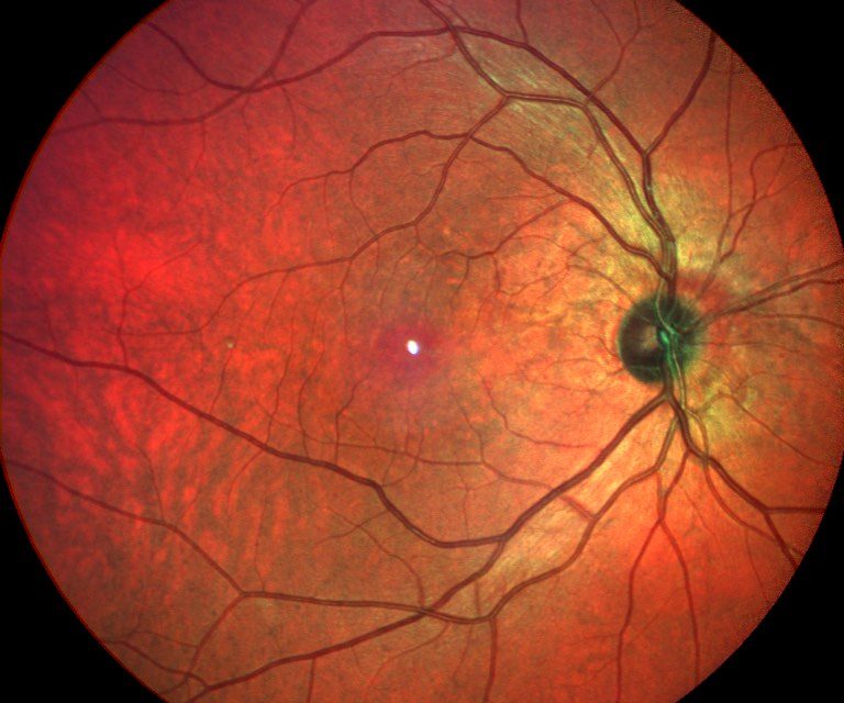
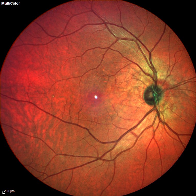
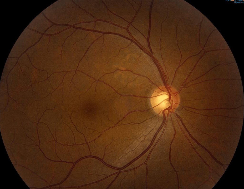
Fundus examination of the right eye (OD) displayed a normal optic disc, macula, and retinal periphery. In contrast, the left eye (OS) exhibited a localized area of retinal elevation and ellipsoid zone disruption at the macula, approximately 1 disc diameter in size, with no associated subretinal fluid or hemorrhage.
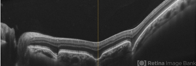
Optical Coherence Tomography (OCT) revealed normal retinal and choroidal anatomy in the right eye (OD) but showed Focal Choroidal Excavation (FCE) corresponding to the area of retinal elevation seen on fundus examination in the left eye (OS), with an ill-defined choroid-sclera interface in the affected region.
Consequently, the patient received a diagnosis of Focal Choroidal Excavation (FCE) in the left eye.
Focal Choroidal Excavation (FCE) DISEASE entity
Focal Choroidal Excavation (FCE) is a newly recognized clinical entity that is often incidentally found and presents asymptomatic or with mildly decreased visual acuity in the affected eye.
It has characteristic findings on optical coherence tomography (OCT) that can be used to distinguish it from other retinal lesions that may appear similar on ophthalmoscopy.
Focal Choroidal Excavation (FCE) was first described by Jampol in 2006 and coined as a term by Margolis in 2011.
It refers to a concavity of the choroid without local scleral changes that are often identified through a characteristic appearance on optical coherence tomography (OCT). This finding usually presents unilaterally and is found incidentally on exams, though exceptions to these patterns have been reported.
The majority of FCEs are thought to be congenital; however, the etiopathology of FCE remains a subject of active investigation.
While most instances of FCE remain relatively stable over time and are managed with close observation, several complications requiring intervention, such as choroidal neovascularization and serous detachment, have been described.
The precise etiopathology of Focal Choroidal Excavation (FCE) remains to date unclear. It has been suggested that the clinical entity may represent a common morphological manifestation of multiple processes, which may be either congenital or secondarily acquired.
Congenital FCE are thought to be mostly stationary and not likely to be vision threatening, while secondary FCE may be related to a variety of conditions — including pachychoroid disease, chorioretinal inflammation, retinal dystrophies, and malignancy — which may require treatment to prevent complications and disease progression.
In one study of patients aged <40 years old, only 3/1697 (0.18%) of eyes examined were found to have an FCE.
Fundus Explorer Pro
Photograph the retinal findings described here with the phone already in your pocket — 22 D optics and built-in illumination in one handheld unit.
From Choroida — the team behind this siteFocal Choroidal Excavation (FCE) MANAGEMENT
In cases of asymptomatic Focal Choroidal Excavation (FCE) without associated choroidal neovascular changes, observation without treatment is recommended.
A slow increase in concavity size on OCT may be observed, but changes to concavity size might also be noted due to unavoidable variability in imaging modality such as slice orientation.
Some authors have postulated that focal scar contraction and maturation may be responsible for the slow progression in concavity size observed in patients over time.
If there is evidence of lesion expansion or scleral thickening, a full evaluation for secondary causes of FCE should be undertaken to determine if any underlying process can be directly treated. For scleral thinning, surgical reinforcement of the sclera can be considered.
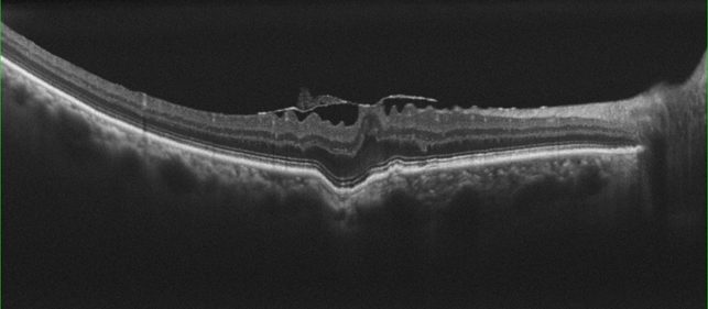
Additional complications may arise that require treatment, including secondary neovascularization and/or serous retinal detachment.
If encountered, these may be managed depending on lesion location and sequelae, with anti-VEGF agents, verteporfin photodynamic therapy, or focal laser photocoagulation.


Document what you see
Two smartphone imaging tools built for everyday clinic use — one for the slit lamp, one for the fundus.
From Choroida — the team behind this siteREFERENCES
- Jampol, L. M. et al. Diagnostic and therapeutic challenges. Retina 26, 1072–1076 (2006).
- Margolis, R. et al. The expanded spectrum of focal choroidal excavation. Arch. Ophthalmol. 129, 1320–1325 (2011).
- Braimah, I. Z., Rapole, S., Dumpala, S. & Chhablani, J. Focal Choroidal Excavation in Retinal Dystrophies. Semin. Ophthalmol. 33, 161–166 (2018).
- Shinojima, A., Kawamura, A., Mori, R. & Yuzawa, M. Morphologic features of focal choroidal excavation on spectral domain optical coherence tomography with simultaneous angiography. Retina 34, 1407–1414 (2014).
- Obata, R. et al. Tomographic and angiographic characteristics of eyes with macular focal choroidal excavation. Retina 33, 1201–1210 (2013).
- Verma, S. et al. Focal choroidal excavation: review of literature. Br. J. Ophthalmol. 105, 1043–1048 (2021).
- Shah, R. C., Gopalakrishnan, M., Goyal, A., Anantharaman, G. & Sethia, A. Focal choroidal excavation: Cause or effect? Indian J. Ophthalmol. 67, 696–698 (2019).
Test yourself
A few questions straight from this article.
-
Focal choroidal excavation refers to which structural change?
FCE is defined as a concavity of the choroid occurring without local scleral change, recognised by its characteristic OCT appearance. -
How does focal choroidal excavation usually present?
FCE is typically a unilateral incidental finding, with patients either asymptomatic or reporting only mildly reduced acuity, though exceptions are reported. -
What makes optical coherence tomography valuable in assessing focal choroidal excavation?
FCE has characteristic OCT features that separate it from other retinal lesions which can appear similar on ophthalmoscopy alone. -
How does congenital focal choroidal excavation typically behave over time?
Congenital FCE lesions are thought to be largely stationary and not likely to threaten vision, in contrast to secondary FCE. -
Which group of conditions has been linked to secondary focal choroidal excavation?
Secondary FCE has been associated with pachychoroid disease, chorioretinal inflammation, retinal dystrophies and malignancy, any of which may need treatment. -
In one study of patients under 40 years of age, how often was focal choroidal excavation found?
Only 3 of 1697 eyes examined in patients under 40, about 0.18%, were found to have a focal choroidal excavation. -
How is asymptomatic focal choroidal excavation without neovascular change managed?
Where FCE is asymptomatic and shows no associated choroidal neovascular change, observation without treatment is recommended. -
Which complications of focal choroidal excavation call for active intervention?
Although most FCE lesions stay stable, secondary choroidal neovascularization and serous retinal detachment are described and require treatment. -
Which treatments are used for neovascular or exudative complications of focal choroidal excavation?
Depending on lesion location and sequelae, these complications are managed with anti-VEGF agents, verteporfin photodynamic therapy or focal laser photocoagulation. -
What has been postulated to explain the slow increase in concavity size in focal choroidal excavation?
Some authors attribute the slow enlargement of the concavity to focal scar contraction and maturation, though imaging variability such as slice orientation also contributes.
