
Retinal vasculitis sheathing describes inflammatory cuffing and exudate along the retinal blood vessels, and because different underlying causes of retinal vasculitis tend to produce recognizably different sheathing patterns and vessel involvement, careful attention to exactly what the sheathing looks like, and which vessels it affects, can meaningfully narrow a differential diagnosis that otherwise spans a wide range of systemic and infectious conditions.
What Sheathing Represents
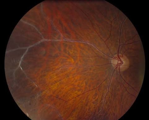
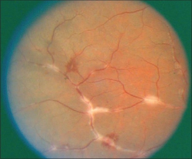
Retinal vasculitis produces perivascular inflammation, visible on fundus examination as a whitish or yellow-white cuffing or sheathing surrounding the affected blood vessel, reflecting inflammatory cell infiltration and exudation around the vessel wall, and the specific character of this sheathing, along with whether it preferentially involves veins, arteries, or both, provides real diagnostic information.
Vein-Predominant Versus Artery-Predominant Involvement
Retinal vasculitis predominantly affecting veins (periphlebitis) is more common overall and is associated with a broad range of causes, including sarcoidosis, multiple sclerosis, and various infectious etiologies, while artery-predominant involvement (periarteritis) is less common and, when present, should raise particular concern for specific causes including Behçet disease and certain infectious retinitis syndromes, including acute retinal necrosis.
Fundus Explorer Pro
Photograph the retinal findings described here with the phone already in your pocket — 22 D optics and built-in illumination in one handheld unit.
From Choroida — the team behind this siteCharacteristic Patterns by Cause
Candle Wax Drippings
Discrete, yellow-white perivascular exudates resembling drips of candle wax along the retinal veins, classically associated with sarcoidosis, and considered one of the more specific fundus findings pointing toward this diagnosis (see phakomatoses and Heerfordt syndrome for a related sarcoidosis presentation).
Frosted Branch Angiitis
A striking pattern of severe, extensive perivascular sheathing affecting both arteries and veins throughout the fundus, giving the retinal vasculature the appearance of frost-covered tree branches, a dramatic finding associated with a range of underlying causes including viral infection (particularly cytomegalovirus, especially in immunocompromised patients), other infectious causes, and, less commonly, an idiopathic or autoimmune presentation.
Segmental Sheathing With Occlusion
Focal, segmental areas of vascular sheathing, sometimes with associated vascular occlusion and adjacent retinal whitening, can be seen in acute retinal necrosis, a severe, rapidly progressive viral retinitis typically caused by herpes simplex or varicella-zoster virus, where the combination of arteritis, necrotizing retinitis, and dense vitritis forms a recognizable, urgent clinical picture.
Peripheral Vasculitis With Occlusion
Peripheral retinal vasculitis with associated vascular occlusion and non-perfusion is a recognized pattern in Behçet disease, often accompanying recurrent episodes of explosive intraocular inflammation and carrying significant risk of ischemic complications, including neovascularization, if not adequately controlled.
Systemic Associations to Consider
- Sarcoidosis, particularly with a candle-wax-dripping pattern
- Behçet disease, particularly with occlusive, artery-involving vasculitis and a history of recurrent oral and genital ulceration
- Multiple sclerosis, which can cause a form of retinal periphlebitis
- Systemic lupus erythematosus and other systemic autoimmune and vasculitic conditions
- Infectious causes, including tuberculosis, syphilis, and viral retinitis syndromes, particularly when the pattern suggests artery involvement or is accompanied by necrotizing retinitis
Diagnostic Evaluation
- Careful, detailed fundus examination and documentation of sheathing pattern, distribution, and vessel type involved
- Fluorescein angiography is valuable for confirming the presence and extent of vasculitis, since vessel wall staining and leakage can be present even when sheathing is subtle on direct examination alone, and for assessing areas of non-perfusion that carry their own management implications
- Systemic evaluation directed by the specific pattern observed and associated clinical features, including targeted laboratory testing, chest imaging when sarcoidosis is suspected, and infectious disease workup when an infectious cause is considered
Management
Management is directed primarily at the underlying systemic or infectious cause once identified, since treating retinal vasculitis effectively generally requires addressing the driving process rather than treating the ocular inflammation in isolation.
- Autoimmune and inflammatory causes are managed with corticosteroids and, for more severe or recurrent disease, steroid-sparing systemic immunosuppression
- Infectious causes, particularly viral retinitis syndromes such as acute retinal necrosis, require urgent antiviral therapy given the potential for rapid, severe visual loss
- Areas of retinal ischemia from occlusive vasculitis may require laser photocoagulation to reduce the risk of neovascular complications
Prognosis
Prognosis depends heavily on the underlying cause, the extent of vascular involvement, and how promptly appropriate treatment is initiated, with infectious causes such as acute retinal necrosis carrying particular urgency given the risk of rapid, severe, and sometimes irreversible vision loss if treatment is delayed.
Careful characterization of the specific sheathing pattern at initial presentation, rather than treating retinal vasculitis as a single, generic diagnosis, supports the faster, more targeted systemic workup that many of these underlying causes require for timely, effective treatment.


Document what you see
Two smartphone imaging tools built for everyday clinic use — one for the slit lamp, one for the fundus.
From Choroida — the team behind this siteReferences
- Rosenbaum JT, Sibley CH, Lin P. Retinal vasculitis. Curr Opin Rheumatol. 2016;28:228-235.
- Walton RC, Ashmore ED. Retinal vasculitis. Curr Opin Ophthalmol. 2003;14:413-419.
- Kleiner RC, Kaplan HJ, Shakin JL, Yannuzzi LA, Crosswell HH Jr, McLean WC Jr. Acute frosted retinal periphlebitis. Am J Ophthalmol. 1988;106:27-34.
- Holland GN. Standard diagnostic criteria for the acute retinal necrosis syndrome. Am J Ophthalmol. 1994;117:663-667.
