Conjunctival follicles versus papillae is one of the most practically useful distinctions in all of external eye examination, since these two conjunctival surface reactions, though they can look superficially similar to an untrained eye, point toward largely opposite categories of underlying cause, viral and chlamydial infection on one side, allergic and some chronic irritative conditions on the other, making correct identification genuinely central to reaching the right diagnosis quickly.
Why This Distinction Matters So Much
A patient with red, irritated eyes and a follicular reaction is, in general, being worked up along a very different diagnostic and management pathway than a patient with the same complaint but a papillary reaction, which is exactly why learning to reliably tell these two patterns apart at the slit lamp is one of the foundational skills of external eye examination.
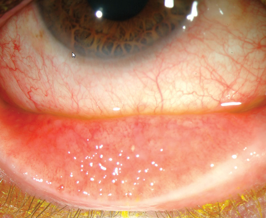
Follicles
Follicles represent focal collections of lymphoid tissue within the substantia propria of the conjunctiva, appearing as multiple small, round, pale, dome-shaped elevations, each typically with tiny blood vessels visible coursing around, rather than through, the individual lesion.
- Most prominent in the inferior fornix and inferior tarsal conjunctiva, though they can occur elsewhere
- Represent a lymphoid, immune-cell-based reaction, classically associated with viral conjunctivitis (particularly adenoviral), chlamydial conjunctivitis (including trachoma and inclusion conjunctivitis), and, notably, chronic topical medication toxicity, which can also provoke a follicular response with sustained use (see keratitis medicamentosa for a related discussion of chronic topical medication effects)
- Generally avascular in their central portion, with the characteristic surrounding, rather than penetrating, vascular pattern
Fundus Explorer Pro
Photograph the retinal findings described here with the phone already in your pocket — 22 D optics and built-in illumination in one handheld unit.
From Choroida — the team behind this sitePapillae
Papillae represent a different structural reaction: a central fibrovascular core surrounded by inflammatory cells, covered by conjunctival epithelium, appearing as a more polygonal, mosaic or cobblestone-like pattern of elevated lesions, each with a visible central vascular tuft, in contrast to the surrounding vessel pattern of a follicle.
- Most prominent on the superior tarsal conjunctiva, in contrast to the inferior predominance typical of follicles
- Represent a vascular, allergic-type reaction, classically associated with allergic conjunctivitis, vernal and atopic keratoconjunctivitis (where giant papillae can form), and contact lens-related giant papillary conjunctivitis
- The central vascular tuft, visible with careful slit-lamp examination, is the key distinguishing structural feature from a follicle
Side-by-Side Comparison
- Location: follicles predominate inferiorly, papillae predominate on the superior tarsal conjunctiva
- Vascular pattern: follicles show vessels surrounding an avascular center; papillae show a central vascular tuft
- Shape: follicles are round and dome-shaped; papillae are more polygonal with a flatter, mosaic top, though giant papillae in severe allergic disease can become quite large and irregular
- Associated cause category: follicles suggest viral or chlamydial infection, or chronic medication toxicity; papillae suggest an allergic or chronic mechanical/contact lens-related process
Using the Distinction Clinically
- A follicular reaction in a patient with acute red eye, watery discharge, and preauricular lymphadenopathy strongly supports viral conjunctivitis, and appropriate counseling about contagion and supportive care follows accordingly
- A follicular reaction that is chronic, rather than acute, particularly with a relevant sexual history or in a neonate, should prompt consideration of chlamydial conjunctivitis
- A papillary reaction with itching as a prominent symptom supports allergic conjunctivitis, while giant papillae in a contact lens wearer point toward contact lens-related giant papillary conjunctivitis, prompting a change in lens care or wear pattern
- Giant papillae combined with limbal findings and a young, atopic patient supports vernal keratoconjunctivitis (see limbal vernal keratoconjunctivitis)
Examination Technique
Eyelid eversion, examining both the superior and inferior tarsal conjunctiva, is essential for a complete assessment, since papillae are most prominent superiorly while follicles are most prominent inferiorly, meaning an examination limited to only the lower lid, or performed without eversion at all, risks missing the very finding most relevant to the correct diagnosis.
Clinical Significance
The follicle-versus-papilla distinction is a foundational piece of external eye examination that, correctly applied, immediately narrows a broad differential diagnosis of red, irritated eyes into one of two largely distinct diagnostic pathways, supporting faster, more targeted, and more accurate subsequent evaluation and management.


Document what you see
Two smartphone imaging tools built for everyday clinic use — one for the slit lamp, one for the fundus.
From Choroida — the team behind this siteReferences
- Leibowitz HM. The red eye. N Engl J Med. 2000;343:345-351.
- Azari AA, Barney NP. Conjunctivitis: a systematic review of diagnosis and treatment. JAMA. 2013;310:1721-1729.
- Bowling B. Kanski’s Clinical Ophthalmology: A Systematic Approach. 8th ed. Edinburgh: Elsevier; 2016.
- Sambursky R, Trattler W, Tauber S, et al. Sensitivity and specificity of the AdenoPlus test for diagnosing adenoviral conjunctivitis. JAMA Ophthalmol. 2013;131:17-22.
Test yourself
A few questions straight from this article.
-
What tissue forms a conjunctival follicle?
Follicles are focal collections of lymphoid tissue within the conjunctival substantia propria, appearing as small, round, pale, dome-shaped elevations. -
What is the structural core of a conjunctival papilla?
A papilla has a central fibrovascular core ringed by inflammatory cells and covered by conjunctival epithelium. -
Which slit-lamp feature identifies a lesion as a papilla rather than a follicle?
The visible central vascular tuft is the key structural difference from a follicle, where vessels course around rather than through the lesion. -
Where are conjunctival follicles most prominent?
Follicles predominate inferiorly, in the inferior fornix and inferior tarsal conjunctiva, although they can occur elsewhere. -
Where are conjunctival papillae most prominent?
Papillae predominate on the superior tarsal conjunctiva, in contrast to the inferior predominance typical of follicles. -
What shape do conjunctival papillae typically take?
Papillae are more polygonal with a flatter mosaic or cobblestone top, while follicles are round and dome-shaped. -
Which non-infectious cause can provoke a conjunctival follicular reaction?
Sustained use of topical medication can itself provoke a follicular response, alongside the viral and chlamydial infectious causes. -
An acute follicular reaction with watery discharge and preauricular lymphadenopathy most supports which diagnosis?
That combination strongly supports viral conjunctivitis, prompting counselling about contagion and supportive care. -
A chronic follicular reaction in a neonate should raise suspicion of which infection?
A follicular reaction that is chronic rather than acute, particularly in a neonate or with a relevant sexual history, should prompt consideration of chlamydial conjunctivitis. -
Which examination step is essential to separate follicles from papillae reliably?
Papillae sit mainly superiorly and follicles mainly inferiorly, so examining only the lower lid, or without eversion, risks missing the decisive finding.

Writes and reviews the clinical articles on Educate. Every post is checked against peer-reviewed sources before it goes live.