
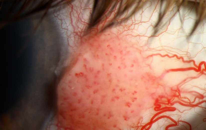
Conjunctival intraepithelial neoplasia sits within a broader category called ocular surface squamous neoplasia, and its clinical appearance can range from subtle to obvious, which is exactly why any persistent, atypical limbal or conjunctival lesion in an older or sun-exposed patient deserves a biopsy rather than an assumption, since distinguishing this premalignant or early malignant process from a benign lesion cannot be done reliably by appearance alone.
What Ocular Surface Squamous Neoplasia Covers
Ocular surface squamous neoplasia is an umbrella term spanning a spectrum from conjunctival intraepithelial neoplasia, in which abnormal squamous cell changes are confined to the epithelium without breaching the basement membrane, through to invasive squamous cell carcinoma, in which the abnormal cells have invaded beyond the epithelium into deeper tissue.
This spectrum concept matters clinically because the appearance of a lesion at the slit lamp does not reliably distinguish where on this spectrum a given lesion actually sits, which is why histopathologic evaluation, not clinical impression alone, determines the definitive diagnosis and grade.
Risk Factors
- Chronic ultraviolet light exposure, the best-established risk factor, explaining the higher incidence in regions of intense sun exposure and in outdoor workers
- Human papillomavirus infection, implicated in a substantial proportion of cases
- HIV infection and other causes of immunosuppression, associated with both higher incidence and a tendency toward more aggressive disease
- Older age, with incidence rising accordingly
- Fair skin and lighter eye color, consistent with the broader pattern of ultraviolet-related risk
All-fit Slit-Lamp Adapter
Record and share exactly what you see at the slit lamp. One adapter fits any slit lamp or surgical microscope — and any smartphone.
From Choroida — the team behind this siteClinical Appearance
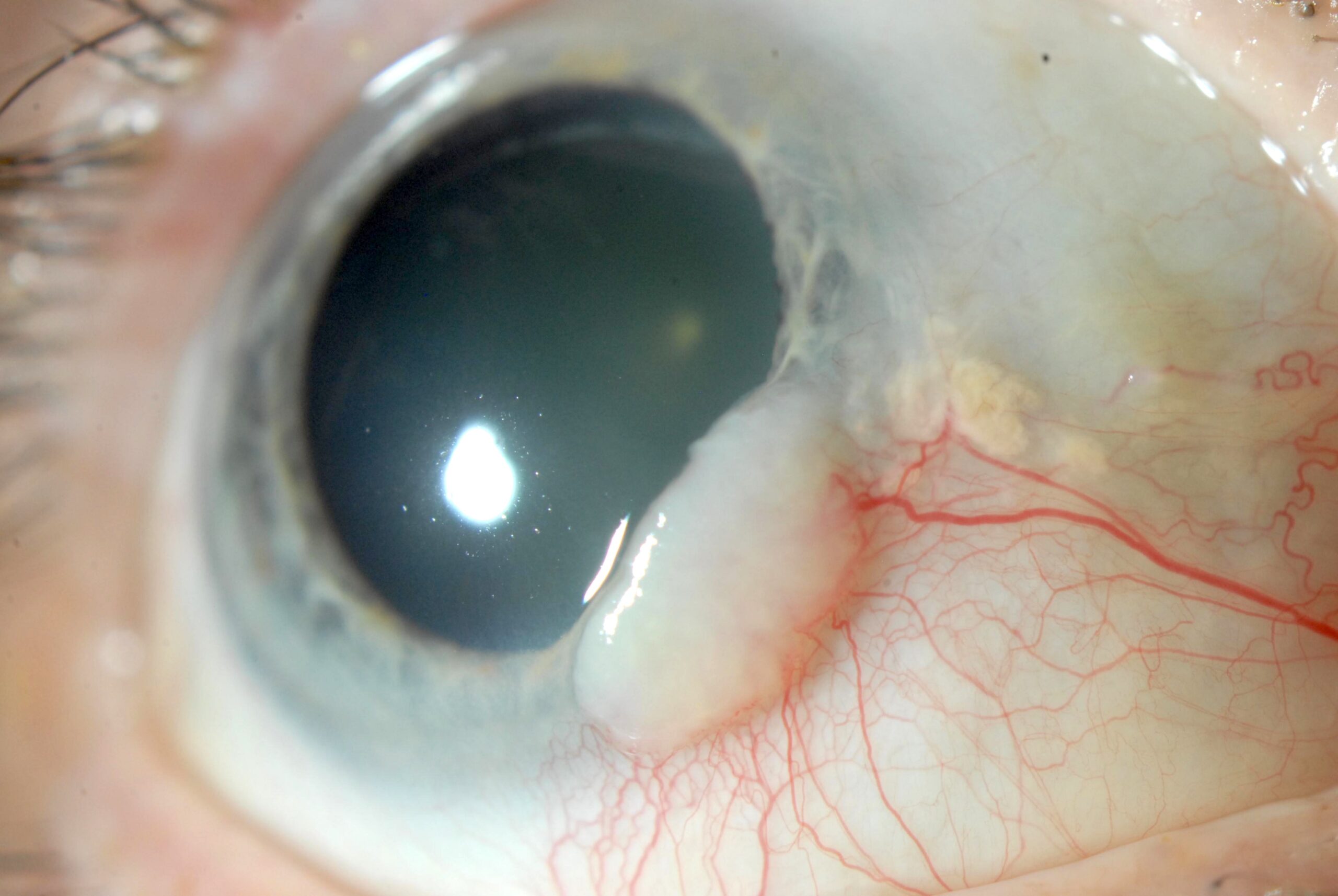
- A gelatinous, fleshy lesion at or near the limbus, often with feeder vessels visible extending into the lesion
- A leukoplakic (white, keratinized) surface can overlie part of the lesion, reflecting surface keratinization
- A papilliform (fronded, papillary) growth pattern in some lesions
- Lesions most commonly arise at or adjacent to the limbus in the interpalpebral zone, corresponding to the area of greatest chronic ultraviolet exposure
- The clinical appearance alone often cannot reliably distinguish conjunctival intraepithelial neoplasia from invasive squamous cell carcinoma or from certain benign mimics, including pterygium or papilloma, reinforcing the need for tissue diagnosis
Diagnostic Evaluation
- Biopsy, either incisional or excisional, provides the definitive diagnosis and grading, and is generally preferred whenever the lesion is amenable to safe removal
- Impression cytology, a less invasive technique sampling superficial cells, can support diagnosis and monitor treatment response in some settings, though it does not provide the same degree of information as a full-thickness biopsy regarding invasion
- High-resolution anterior segment optical coherence tomography is increasingly used as a non-invasive adjunct, helping characterize epithelial thickening and supporting monitoring, particularly useful for following a lesion over time without repeated biopsy
- A careful, wide-field slit-lamp examination of the entire ocular surface, since lesions can occasionally be multifocal
Management
Surgical Excision
Complete surgical excision with a clear margin, often combined with adjunctive cryotherapy to the surrounding conjunctiva to reduce local recurrence risk, is a standard, effective approach for localized, resectable lesions, using a “no-touch” technique to minimize the risk of seeding tumor cells to adjacent tissue during removal.
Topical Chemotherapy
- Topical mitomycin C, applied as eye drops in cycles, is effective for treating conjunctival intraepithelial neoplasia, either as primary treatment for more diffuse lesions not ideally suited to excision, or as adjunctive treatment after surgery to reduce recurrence
- Topical interferon alpha-2b has also shown good efficacy with a generally more favorable side-effect profile than mitomycin C, making it an appealing option particularly for larger or diffuse lesions, though treatment courses are typically longer
- 5-fluorouracil is another topical chemotherapeutic option used in some treatment protocols
Managing Invasive Disease
When biopsy reveals invasive squamous cell carcinoma rather than disease confined to the epithelium, more extensive surgical excision is needed, and in rare cases with orbital invasion, more extensive surgery, including orbital exenteration, may be required.
Follow-Up
Given a meaningful risk of local recurrence even after apparently complete treatment, regular, sustained follow-up examination of the treated area and the broader ocular surface is an essential part of long-term management, continuing for years after apparently successful initial treatment.
Prognosis
With appropriate treatment, whether surgical, topical chemotherapeutic, or a combination, conjunctival intraepithelial neoplasia generally has a good prognosis, and vision and globe preservation are achieved in the great majority of patients when the disease is caught and treated before significant invasive progression.
Delayed diagnosis, allowing progression to more invasive or extensive disease, worsens both the complexity of treatment needed and the overall prognosis, underscoring the value of biopsying persistent or atypical limbal and conjunctival lesions rather than assuming a benign cause based on appearance alone.


Document what you see
Two smartphone imaging tools built for everyday clinic use — one for the slit lamp, one for the fundus.
From Choroida — the team behind this siteReferences
- Lee GA, Hirst LW. Ocular surface squamous neoplasia. Surv Ophthalmol. 1995;39:429-450.
- Shields CL, Shields JA. Tumors of the conjunctiva and cornea. Surv Ophthalmol. 2004;49:3-24.
- Nanji AA, Sayyad FE, Galor A, Dubovy S, Karp CL. High-resolution optical coherence tomography as an adjunctive tool in the diagnosis of corneal and conjunctival pathology. Ocul Surf. 2015;13:226-235.
- Shields CL, Kaliki S, Kim HJ, et al. Interferon for ocular surface squamous neoplasia in 81 cases: outcomes based on the American Joint Committee on Cancer classification. Cornea. 2013;32:248-256.
Test yourself
A few questions straight from this article.
-
What defines conjunctival intraepithelial neoplasia histologically?
In conjunctival intraepithelial neoplasia the abnormal squamous cell changes are confined to the epithelium without breaching the basement membrane, in contrast to invasive squamous cell carcinoma. -
Why is tissue diagnosis rather than slit-lamp impression required for a suspected conjunctival intraepithelial neoplasia?
The slit-lamp appearance does not reliably distinguish where on the squamous neoplasia spectrum a lesion sits, so histopathologic evaluation rather than clinical impression determines the definitive diagnosis. -
What is the best-established risk factor for ocular surface squamous neoplasia?
Chronic ultraviolet exposure is the best-established risk factor, explaining the higher incidence in regions of intense sun exposure and among outdoor workers. -
Which viral infection is implicated in a substantial proportion of ocular surface squamous neoplasia cases?
Human papillomavirus infection is implicated in a substantial proportion of cases, alongside ultraviolet exposure and immunosuppression as recognised risk factors. -
What is the typical clinical appearance of a conjunctival intraepithelial neoplasia lesion?
The lesion is typically gelatinous and fleshy at or near the limbus, often with visible feeder vessels, and may carry a leukoplakic keratinised surface or a papilliform growth pattern. -
Which benign lesions can mimic conjunctival intraepithelial neoplasia on appearance alone?
Clinical appearance often cannot reliably separate conjunctival intraepithelial neoplasia from invasive carcinoma or from benign mimics including pterygium and papilloma, reinforcing the need for tissue diagnosis. -
What role does high-resolution anterior segment OCT play in conjunctival intraepithelial neoplasia?
High-resolution anterior segment OCT is increasingly used as a non-invasive adjunct that characterises epithelial thickening and supports following a lesion over time without repeated biopsy. -
Why is a no-touch technique used when excising conjunctival intraepithelial neoplasia?
Complete excision with a clear margin uses a no-touch technique to minimise the risk of seeding tumour cells to adjacent tissue during removal, often combined with adjunctive cryotherapy to reduce local recurrence. -
How does topical interferon alpha-2b compare with mitomycin C for conjunctival intraepithelial neoplasia?
Topical interferon alpha-2b shows good efficacy with a generally more favourable side-effect profile than mitomycin C, making it appealing for larger or diffuse lesions, though treatment courses are typically longer. -
What follow-up is required after apparently complete treatment of conjunctival intraepithelial neoplasia?
Because there is a meaningful risk of local recurrence even after apparently complete treatment, regular examination of the treated area and the broader ocular surface continues for years.

Writes and reviews the clinical articles on Educate. Every post is checked against peer-reviewed sources before it goes live.


