
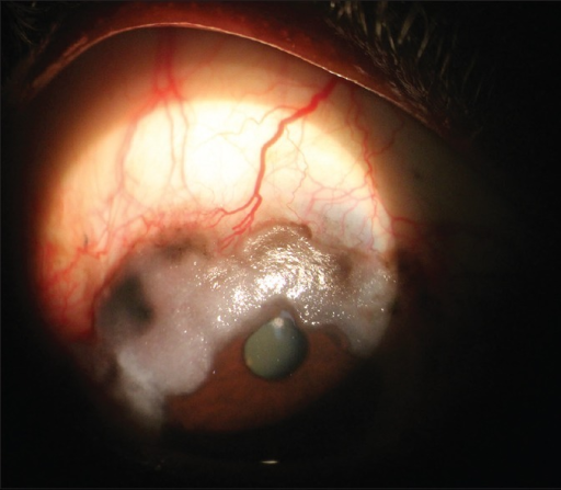
Conjunctival lymphoma typically presents as a distinctive, soft, salmon-pink or salmon-colored mass beneath the conjunctiva, and while the ocular finding itself is often readily treatable, its real clinical significance lies in what it can represent systemically, since a meaningful proportion of patients have, or will develop, lymphoma at another site in the body, making systemic staging a mandatory rather than optional part of the workup.
Clinical Presentation
- A characteristic soft, mobile, salmon-colored subconjunctival mass, most often located in the fornix or bulbar conjunctiva, though it can occur elsewhere on the ocular surface
- The lesion is generally painless and slow-growing, and patients often present because of a visible or palpable mass rather than significant discomfort
- Can be unilateral or bilateral
- Growth over weeks to months is typical, distinguishing it from the more acute presentation of an infectious or inflammatory conjunctival process
Types of Conjunctival Lymphoma
The majority of conjunctival lymphomas are low-grade, B-cell non-Hodgkin lymphomas, most commonly extranodal marginal zone lymphoma of mucosa-associated lymphoid tissue (MALT lymphoma), which tends to behave relatively indolently.
Less commonly, higher-grade lymphomas, including diffuse large B-cell lymphoma, can present in the conjunctiva, and these carry a different, generally more aggressive natural history and treatment approach, which is part of why histologic confirmation and subtyping through biopsy is essential rather than assuming a uniform, indolent course for every conjunctival lymphoma.
All-fit Slit-Lamp Adapter
Record and share exactly what you see at the slit lamp. One adapter fits any slit lamp or surgical microscope — and any smartphone.
From Choroida — the team behind this siteDiagnostic Evaluation
Biopsy
Incisional or excisional biopsy of the conjunctival mass is necessary for diagnosis, with the tissue sent for histopathology, immunohistochemistry, and flow cytometry to establish the specific lymphoma subtype, since management and prognosis differ meaningfully between low-grade and high-grade lymphomas.
Systemic Staging
Once conjunctival lymphoma is confirmed, systemic staging is essential, since ocular adnexal lymphoma, including conjunctival lymphoma, can be an isolated finding, a presenting sign of previously undiagnosed systemic lymphoma, or evidence of recurrence in a patient with a known lymphoma history.
- Physical examination for lymphadenopathy and organomegaly
- Systemic imaging, typically CT or PET-CT, to evaluate for lymphoma elsewhere in the body
- Bone marrow biopsy is considered in some cases, depending on the lymphoma subtype and other clinical findings
- Coordinated evaluation with hematology-oncology for complete staging and to determine whether the conjunctival finding represents isolated ocular disease or a manifestation of systemic lymphoma
Management
Localized, Low-Grade Disease
For conjunctival lymphoma confirmed to be localized, without evidence of systemic involvement, treatment options include local radiotherapy, which is highly effective for MALT lymphoma confined to the conjunctiva, and, in select cases, observation or local excision alone for very small, indolent lesions.
Systemic Disease
When systemic lymphoma is identified, whether the conjunctival lesion is the presenting sign or a manifestation of known disease, management is coordinated with hematology-oncology and follows systemic lymphoma treatment protocols appropriate to the specific subtype and stage, with the conjunctival component addressed within that broader systemic treatment plan.
Radiotherapy Considerations
Local radiotherapy to the conjunctiva and ocular adnexa is generally well tolerated and effective, though it carries a risk of certain ocular side effects, including dry eye and, less commonly, cataract, which are discussed with patients as part of treatment planning.
Prognosis
Low-grade, localized conjunctival MALT lymphoma generally has an excellent prognosis, with high rates of local control following radiotherapy and good long-term outcomes, particularly when systemic staging confirms truly isolated disease.
Higher-grade lymphoma subtypes and cases associated with systemic lymphoma involvement carry a prognosis that depends primarily on the systemic disease characteristics and its response to systemic treatment, reinforcing why the conjunctival finding, however reassuring it may appear locally, must always be interpreted in the context of a complete systemic evaluation.


Document what you see
Two smartphone imaging tools built for everyday clinic use — one for the slit lamp, one for the fundus.
From Choroida — the team behind this siteReferences
- Coupland SE, Krause L, Delecluse HJ, et al. Lymphoproliferative lesions of the ocular adnexa: analysis of 112 cases. Ophthalmology. 1998;105:1430-1441.
- Shields CL, Shields JA, Carvalho C, Rundle P, Smith AF. Conjunctival lymphoid tumors: clinical analysis of 117 cases and relationship to systemic lymphoma. Ophthalmology. 2001;108:979-984.
- Sjo LD. Ophthalmic lymphoma: epidemiology and pathogenesis. Acta Ophthalmol. 2009;87:1-20.
- Stafford SL, Kozelsky TF, Garrity JA, et al. Orbital lymphoma: radiotherapy outcome and complications. Radiother Oncol. 2001;59:139-144.
