Conjunctival lymphoma is a malignant growth of lymphocytes in the conjunctiva, and it usually appears as a painless, smooth, salmon-pink mass under the surface of the eye. Most cases are low-grade B-cell tumors that respond well to local treatment. Because some patients have or later develop lymphoma elsewhere, every case needs a biopsy and full systemic staging.
Conjunctival Lymphoma Symptoms and Signs
Most patients notice a pink, fleshy lump, mild irritation, or a foreign body sensation, and many have no pain at all. The mass grows slowly over weeks to months, which separates it from acute infectious or allergic conjunctivitis. Vision stays normal unless a large lesion disturbs the tear film or limits eye movement.
The typical patient is an older adult, and women are affected slightly more often than men. Examination usually shows the following:
- A soft, mobile subconjunctival mass in the fornix or on the bulbar conjunctiva, sometimes reaching the limbus
- A smooth surface with fine overlying vessels and intact epithelium
- Unilateral disease in most patients, though bilateral lesions occur
- Mild redness or tearing, and ptosis or restricted motility if the tumor extends into the lid or orbit
What Conjunctival Lymphoma Looks Like: Pictures and the Salmon Patch
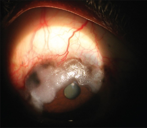
In clinical photographs of conjunctiva lymphoma, the lesion appears as a diffuse, fleshy, salmon-colored elevation that molds to the globe and fornix instead of forming a discrete cyst. The salmon patch of the conjunctiva reflects a dense, uniform infiltrate of lymphocytes with fine vessels running through it. The patch may be flat or form a thick fold.
Color alone never proves the diagnosis. Anterior segment OCT typically shows a homogeneous subepithelial mass under intact epithelium, which helps separate it from epithelial tumors, but only tissue confirms lymphoma. Photograph the lesion at baseline so you can compare size after treatment.
All-fit Slit-Lamp Adapter
Record and share exactly what you see at the slit lamp. One adapter fits any slit lamp or surgical microscope — and any smartphone.
From Choroida — the team behind this siteConjunctival MALT Lymphoma and Other Subtypes
Most conjunctival lymphomas are extranodal marginal zone lymphomas of mucosa-associated lymphoid tissue, called conjunctival MALT lymphoma. These low-grade B-cell non-Hodgkin tumors behave indolently and often stay localized for a long time.
Less often, follicular lymphoma, mantle cell lymphoma, or diffuse large B-cell lymphoma presents in the conjunctiva. These subtypes carry a different and sometimes more aggressive course, so biopsy with subtyping is essential. Do not assume that every salmon-colored lesion follows the indolent pattern.
What Causes Lymphoma of the Conjunctiva?
The exact cause is unknown, but chronic antigenic stimulation is the leading explanation, which links these tumors to autoimmune disease such as Sjögren syndrome. Chlamydia psittaci has been found in some ocular adnexal lymphomas, with marked geographic variation. Most patients are older than 50.
Conjunctival Lymphoma Differential Diagnosis
Several lesions mimic a salmon-colored conjunctival mass, and clinical appearance cannot separate them reliably. The main entities to consider are:
- Reactive lymphoid hyperplasia: looks identical and needs histology plus clonality testing to distinguish
- Ocular surface squamous neoplasia: usually gelatinous or leukoplakic at the limbus, as in conjunctival intraepithelial neoplasia
- Amelanotic melanoma: a pigment-free pink lesion that is often more vascular and firmer
- Pyogenic granuloma: red, friable, and typically after surgery or trauma
- Dermolipoma: a yellow-white mass at the lateral canthus, sometimes with hairs
- Amyloidosis, sarcoidosis, lymphangioma, and Kaposi sarcoma: less common mimics, with Kaposi sarcoma mainly in immunocompromised patients
A smooth, painless, slowly enlarging salmon lesion favors lymphoma, while pigment, keratinization, or a friable surface points elsewhere. Reactive lymphoid hyperplasia is the hardest to exclude, which is another reason every suspicious lesion goes to the pathologist.
How Doctors Diagnose Conjunctival Lymphoma
Before biopsy, measure and photograph the lesion, evert the lids to see the fornices, and palpate the orbit and the preauricular and cervical nodes. Ask about prior lymphoma, autoimmune disease, immunosuppression, and systemic symptoms. These steps take minutes and shape how aggressively you stage.
Incisional or excisional biopsy is required. Send part of the specimen fresh rather than fixed in formalin, so the pathologist can run flow cytometry alongside histology, immunohistochemistry, and molecular clonality studies. Handle the tissue gently, because lymphoid cells crush easily. These tests establish the subtype, and subtype drives both treatment and prognosis.
Why Conjunctival Lymphoma Needs Systemic Staging
Ocular adnexal lymphoma can be an isolated finding, the first sign of undiagnosed systemic lymphoma, or a recurrence in a patient with known disease. A meaningful proportion of patients have involvement elsewhere at diagnosis or develop it later, so staging is part of the standard workup. Coordinate it with hematology-oncology.
- Physical examination for lymphadenopathy and hepatosplenomegaly
- CT or PET-CT of the neck, chest, abdomen, and pelvis
- Laboratory tests, including complete blood count, lactate dehydrogenase, and hepatitis B screening before rituximab
- Bone marrow biopsy in selected cases, depending on subtype and findings
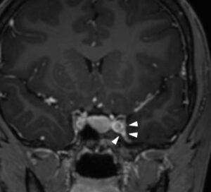
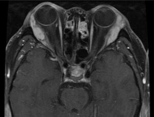
- Orbital MRI when the tumor extends beyond the conjunctiva
Conjunctival Lymphoma Treatment
For localized low-grade disease, low-dose external beam radiotherapy gives excellent local control and is the usual first choice. Very small, indolent lesions may be observed or excised, and rituximab or other systemic therapy can be used when radiation is unsuitable. Some centers try doxycycline when Chlamydia psittaci is detected, with variable results.
When staging finds systemic lymphoma, hematology-oncology directs treatment according to subtype and stage, and the conjunctival tumor is treated within that plan. High-grade lymphomas such as diffuse large B-cell lymphoma usually need chemoimmunotherapy rather than local treatment alone.
Treatment choice also reflects patient factors. Age, comorbidity, bilateral disease, and eyelid or orbital extension all influence whether radiation, systemic therapy, or observation fits best, and the decision is shared among ophthalmology, radiation oncology, and hematology-oncology.
Radiotherapy is generally well tolerated, but it can cause dry eye and, less often, cataract, keratopathy, or radiation retinopathy. Shielding and dose planning reduce these risks, and you should discuss them before treatment. Lubricants and regular follow-up manage most ocular surface effects.
Prognosis and Follow-Up
Localized, low-grade conjunctival MALT lymphoma has an excellent prognosis, with high rates of local control after radiotherapy and good long-term survival. Recurrence can still appear in the other eye, in other ocular adnexal sites, or in distant extranodal tissue, so follow-up continues over the long term.
After treatment, examine the ocular surface and fornices at regular intervals, and repeat imaging as oncology directs. Ask about new lumps, night sweats, weight loss, or fevers, and examine the fellow eye and lids. Refer promptly if the lesion regrows.
Higher-grade subtypes and cases with systemic involvement depend mainly on the systemic disease and its response to treatment. A reassuring local appearance therefore never replaces a complete systemic evaluation. Lymphoma inside the eye is a separate problem, covered in our review of intraocular lymphoma management strategies.


Document what you see
Two smartphone imaging tools built for everyday clinic use — one for the slit lamp, one for the fundus.
From Choroida — the team behind this siteReferences
- Coupland SE, Krause L, Delecluse HJ, et al. Lymphoproliferative lesions of the ocular adnexa: analysis of 112 cases. Ophthalmology. 1998;105:1430-1441.
- Shields CL, Shields JA, Carvalho C, Rundle P, Smith AF. Conjunctival lymphoid tumors: clinical analysis of 117 cases and relationship to systemic lymphoma. Ophthalmology. 2001;108:979-984.
- Sjo LD. Ophthalmic lymphoma: epidemiology and pathogenesis. Acta Ophthalmol. 2009;87:1-20.
- Stafford SL, Kozelsky TF, Garrity JA, et al. Orbital lymphoma: radiotherapy outcome and complications. Radiother Oncol. 2001;59:139-144.
Test yourself
A few questions straight from this article.
-
Which appearance is characteristic of conjunctival lymphoma?
The classic lesion is a soft, mobile, salmon-coloured mass beneath the conjunctiva, most often in the fornix or bulbar conjunctiva. -
Which growth pattern helps separate conjunctival lymphoma from an infectious or inflammatory conjunctival process?
Conjunctival lymphoma is generally painless and slow-growing over weeks to months, unlike the more acute presentation of an infectious or inflammatory conjunctival process. -
Which lymphoma subtype accounts for the majority of conjunctival lymphomas?
Most conjunctival lymphomas are low-grade B-cell non-Hodgkin lymphomas, most commonly extranodal marginal zone lymphoma of mucosa-associated lymphoid tissue, which behaves relatively indolently. -
Which higher-grade lymphoma can present in the conjunctiva with a more aggressive natural history?
Less commonly, higher-grade lymphomas such as diffuse large B-cell lymphoma arise in the conjunctiva and carry a generally more aggressive course and different treatment approach. -
Why is histologic subtyping of a conjunctival lymphoma essential rather than assuming an indolent course?
Biopsy with histopathology, immunohistochemistry and flow cytometry establishes the subtype because management and prognosis differ meaningfully between low-grade and high-grade lymphomas. -
Which imaging is typically used for systemic staging once conjunctival lymphoma is confirmed?
Systemic staging typically uses CT or PET-CT to look for lymphoma elsewhere, alongside examination for lymphadenopathy and organomegaly. -
Why is systemic staging mandatory rather than optional in conjunctival lymphoma?
Ocular adnexal lymphoma can be isolated, the presenting sign of previously undiagnosed systemic lymphoma, or evidence of recurrence in someone with a known lymphoma history. -
What is the treatment of choice for MALT lymphoma confirmed to be confined to the conjunctiva?
Local radiotherapy is highly effective for MALT lymphoma confined to the conjunctiva, with observation or local excision alone reserved for very small, indolent lesions. -
Which ocular side effects are recognised after local radiotherapy to the conjunctiva and ocular adnexa?
Local radiotherapy is generally well tolerated and effective but carries a risk of dry eye and, less commonly, cataract, discussed during treatment planning. -
What is the outlook for localised, low-grade conjunctival MALT lymphoma?
Low-grade localised conjunctival MALT lymphoma generally has an excellent prognosis with high local control after radiotherapy, particularly when staging confirms truly isolated disease.

Writes and reviews the clinical articles on Educate. Every post is checked against peer-reviewed sources before it goes live.