
Atopic keratoconjunctivitis is a chronic, bilateral allergic ocular surface disease occurring in patients with atopic dermatitis, and its most feared complication, a corneal shield ulcer, results not from the allergic inflammation directly but from the mechanical trauma of the relentless eye rubbing that severe itching drives, which is why controlling the itch is as central to preventing the ulcer as controlling the inflammation itself.
How Atopic Keratoconjunctivitis Differs From Simpler Allergic Conjunctivitis
Unlike seasonal or perennial allergic conjunctivitis, which is typically self-limited and rarely threatens vision, atopic keratoconjunctivitis is a chronic, often year-round condition affecting patients with underlying atopic dermatitis, and it carries a real risk of corneal scarring, vascularization, and vision loss if inadequately controlled over years.
It shares some features with vernal keratoconjunctivitis, another severe allergic eye disease, but atopic keratoconjunctivitis tends to affect an older age group, persists into and through adulthood rather than resolving after puberty, and is more closely tied to eyelid skin disease.
Clinical Features
- Chronic bilateral itching, redness, and mucoid discharge, typically worse than in simple allergic conjunctivitis and less clearly seasonal
- Eyelid skin changes reflecting the underlying atopic dermatitis: thickened, lichenified, sometimes fissured skin, with a tendency toward secondary staphylococcal blepharitis
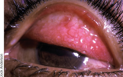
- Papillary conjunctival reaction, generally less florid than the giant papillae typical of vernal keratoconjunctivitis but still contributing to surface irritation
- Corneal involvement ranging from punctate epithelial erosions to, in severe cases, a shield ulcer
- Associated findings can include keratoconus, cataract (including a characteristic anterior subcapsular pattern), and increased risk of herpes simplex keratitis and other infections given the chronic ocular surface compromise
All-fit Slit-Lamp Adapter
Record and share exactly what you see at the slit lamp. One adapter fits any slit lamp or surgical microscope — and any smartphone.
From Choroida — the team behind this siteThe Shield Ulcer
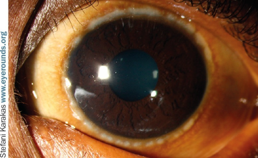
A shield ulcer is an oval or shield-shaped, often superior, sterile corneal epithelial defect with an associated plaque, resulting from mechanical trauma of chronic, vigorous eye rubbing against inflamed, papillae-laden tarsal conjunctiva, compounded by the toxic effects of prolonged inflammatory mediator exposure on the corneal surface.
- Presents with pain, photophobia, and reduced vision
- The base of the ulcer often develops a plaque of altered mucus and inflammatory debris that can impede re-epithelialization if not addressed
- Left untreated, it can lead to permanent corneal scarring within the visual axis
Management
Controlling the Underlying Allergic Disease
- Topical antihistamine and mast cell stabilizer combination drops for ongoing maintenance control
- Topical corticosteroids for acute flares and more severe inflammation, used for limited courses given the cataract and glaucoma risks of chronic steroid use
- Topical calcineurin inhibitors, such as tacrolimus ointment applied to the eyelid skin and, in some regimens, topical cyclosporine or tacrolimus drops for the ocular surface itself, valuable for longer-term control with less steroid-related risk
- Systemic management of the underlying atopic dermatitis, coordinated with dermatology, since better control of the skin disease generally improves the ocular disease as well
Breaking the Rubbing Cycle
Patient education about the mechanical role of eye rubbing in driving both symptoms and complications is central to management, alongside cool compresses and adequate control of the underlying itch with the medical measures above, since asking a patient simply to stop rubbing without addressing the itch that drives it is rarely effective on its own.
Treating a Shield Ulcer
- Aggressive control of the underlying inflammation, as above
- Debridement of the associated plaque when present, to allow the epithelium to heal
- A bandage contact lens to protect the healing epithelium and physically limit further rubbing trauma
- Lubrication and, when needed, a short course of topical antibiotics for prophylaxis against secondary infection while the epithelium is compromised
Prognosis
With adequate long-term control of the underlying allergic and dermatologic disease, most patients avoid the most severe complications, though atopic keratoconjunctivitis tends to be a lifelong condition requiring sustained management rather than a problem that resolves on its own.
A shield ulcer that has caused visual axis scarring may leave permanent visual impairment even after the acute episode resolves, which underscores the importance of early, adequate control of the underlying disease before this complication develops.


Document what you see
Two smartphone imaging tools built for everyday clinic use — one for the slit lamp, one for the fundus.
From Choroida — the team behind this siteReferences
- Bielory L. Allergic and immunologic disorders of the eye. Part II: Ocular allergy. J Allergy Clin Immunol. 2000;106:1019-1032.
- Foster CS, Calonge M. Atopic keratoconjunctivitis. Ophthalmology. 1990;97:992-1000.
- Tuft SJ, Kemeny DM, Dart JK, Buckley RJ. Clinical features of atopic keratoconjunctivitis. Ophthalmology. 1991;98:150-158.
- Trocme SD, Sra KK. Spectrum of ocular allergy. Curr Opin Allergy Clin Immunol. 2002;2:423-427.
