
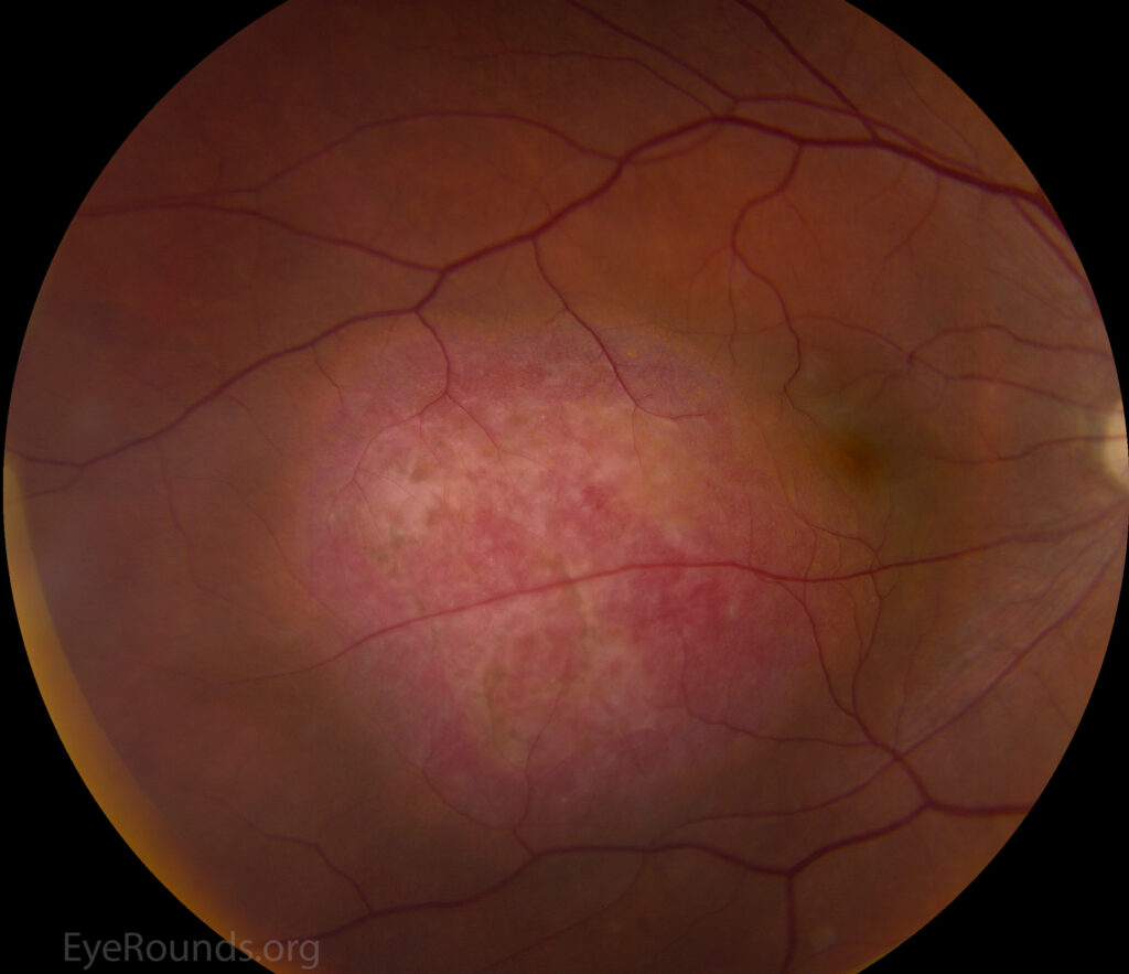
Choroidal hemangioma is a benign vascular tumor of the choroid that comes in two clinically distinct forms — circumscribed and diffuse — with different associations, different presentations, and different management priorities.
Both are congenital vascular hamartomas rather than true neoplasms, though their clinical behavior diverges enough that they are best thought of as separate entities sharing a name, and recognizing which form is present shapes both the systemic workup and the expected long-term course.
Circumscribed Choroidal Hemangioma
The circumscribed form is an isolated, sporadic lesion, most often located near the posterior pole, usually discovered in adulthood when it causes visual symptoms from associated subretinal fluid or, less often, as an incidental finding on routine fundus exam.
It is not associated with any systemic syndrome and occurs as an isolated ocular finding in an otherwise healthy patient.
On fundus exam it appears as an orange-red, dome-shaped, well-circumscribed choroidal mass, and its color is one of the more distinctive features that helps separate it from other choroidal tumors that can otherwise mimic it, particularly amelanotic choroidal melanoma.
Diffuse Choroidal Hemangioma and Sturge-Weber Syndrome
The diffuse form is strongly associated with Sturge-Weber syndrome, where it accompanies the facial port-wine stain and leptomeningeal vascular malformation that define the syndrome.
Rather than a discrete mass, the diffuse form thickens the choroid over a much wider area, sometimes described as giving the fundus a “tomato ketchup” appearance from the diffusely thickened, deep red choroid visible through the retina.
Because the diffuse form is part of a recognized syndrome, its presence should prompt confirmation of the other features of Sturge-Weber syndrome if not already established, and coordination with the broader care already in place for that diagnosis, including glaucoma surveillance, since ipsilateral glaucoma is common in Sturge-Weber syndrome independent of the choroidal finding itself.
Because the facial port-wine stain is typically present from birth and highly visible, the diffuse choroidal hemangioma is usually identified during the ophthalmic workup that follows a Sturge-Weber diagnosis already made on dermatologic grounds, rather than being the finding that leads to the diagnosis in the first place.
Fundus Explorer Pro
Photograph the retinal findings described here with the phone already in your pocket — 22 D optics and built-in illumination in one handheld unit.
From Choroida — the team behind this siteClinical Presentation
Circumscribed lesions are frequently asymptomatic when small and peripheral but cause metamorphopsia, blurred vision, or a visual field defect when they involve or approach the macula, largely through the exudative subretinal fluid they can generate over time rather than through the mass itself.
Diffuse lesions in Sturge-Weber syndrome often present earlier, in childhood, sometimes with a shallow, chronic exudative retinal detachment that can be subtle and easy to miss without a careful, dilated exam.
Because a young child cannot always reliably report visual symptoms, and because the exudative detachment associated with diffuse disease can be gradual and asymmetric between the two eyes, a formal dilated fundus exam at the time Sturge-Weber syndrome is first diagnosed — rather than waiting for a specific visual complaint — is the practical way this finding is usually caught early.
Differential Diagnosis
- Amelanotic choroidal melanoma — the most important alternative diagnosis to exclude, given the difference in management and prognosis
- Choroidal metastasis — typically less well-circumscribed, with a relevant primary malignancy history
- Choroidal osteoma — calcified, seen on ultrasound or CT, more common in young women
- Posterior scleritis — inflammatory, painful, associated with other signs of scleral inflammation
Distinguishing circumscribed choroidal hemangioma from amelanotic melanoma is the single most consequential differential decision, and it relies heavily on multimodal imaging rather than fundus appearance alone, since both can present as an orange to reddish elevated posterior pole mass on routine fundus exam.
Diagnostic Evaluation
B-scan ultrasonography shows a characteristically high internal reflectivity for a choroidal hemangioma, in contrast to the low-to-medium reflectivity typical of choroidal melanoma, making ultrasound one of the more discriminating tests available.
Indocyanine green angiography classically shows early, rapid hyperfluorescence of the tumor’s intrinsic vasculature followed by a “washout” phenomenon in later phases, a pattern that is distinctive enough to be considered strongly supportive of the diagnosis when present.
OCT is used to characterize and monitor any associated subretinal fluid, which is often the more clinically relevant driver of vision loss and treatment decisions rather than the size of the tumor itself.
Management
Asymptomatic, small circumscribed lesions without subretinal fluid can simply be observed with periodic monitoring.
Symptomatic lesions with vision-threatening subretinal fluid are treated to induce tumor regression and resolve the fluid: photodynamic therapy has become a preferred option for many circumscribed lesions given its selective effect on the abnormal vasculature, with argon laser photocoagulation and external beam or plaque radiotherapy used in select cases, for larger or diffuse lesions less amenable to photodynamic therapy.
Diffuse hemangiomas in Sturge-Weber syndrome are managed similarly when symptomatic, with the added consideration of coordinating care around the broader syndrome, including glaucoma control, which can itself affect visual outcomes independent of the choroidal lesion.
Because glaucoma in Sturge-Weber syndrome can develop insidiously and contribute to visual loss just as significantly as the choroidal hemangioma itself, regular intraocular pressure monitoring should continue as a permanent part of these patients’ ophthalmic care, not just during periods when the choroidal lesion itself is being actively managed.


Document what you see
Two smartphone imaging tools built for everyday clinic use — one for the slit lamp, one for the fundus.
From Choroida — the team behind this siteReferences
- Shields CL, Honavar SG, Shields JA, et al. Circumscribed choroidal hemangioma: clinical manifestations and factors predictive of visual outcome. Ophthalmology.
- Witschel H, Font RL. Hemangioma of the choroid: a clinicopathologic study of 71 cases. Survey of Ophthalmology.
- Sullivan TJ, Clarke MP, Morin JD. The ocular manifestations of the Sturge-Weber syndrome. Journal of Pediatric Ophthalmology and Strabismus.
- American Academy of Ophthalmology. Basic and Clinical Science Course, Section 4: Ophthalmic Pathology and Intraocular Tumors.
Test yourself
A few questions straight from this article.
-
Choroidal hemangiomas are best classified as which type of lesion?
Both the circumscribed and diffuse forms are congenital vascular hamartomas rather than true neoplasms, which is why they behave as benign, non-progressive vascular lesions. -
Diffuse choroidal hemangioma is strongly associated with which systemic syndrome?
The diffuse form accompanies the facial port-wine stain and leptomeningeal vascular malformation that define Sturge-Weber syndrome, so finding it should prompt confirmation of the syndrome's other features. -
How does circumscribed choroidal hemangioma appear on fundus examination?
The orange-red colour of this dome-shaped, sharply demarcated choroidal mass is one of its most distinctive features and helps separate it from other choroidal tumours. -
Which fundus appearance is classically described in diffuse choroidal hemangioma?
Instead of a discrete mass, the diffuse form thickens the choroid over a wide area, and the deep red thickened choroid seen through the retina gives the described "tomato ketchup" appearance. -
What does B-scan ultrasonography characteristically show in choroidal hemangioma?
Choroidal hemangioma shows high internal reflectivity, in contrast to the low-to-medium reflectivity typical of choroidal melanoma, making ultrasound one of the more discriminating tests available. -
What is the classic indocyanine green angiography pattern of choroidal hemangioma?
ICGA shows early, rapid hyperfluorescence of the tumour's intrinsic vasculature with a later "washout" phenomenon, a pattern distinctive enough to strongly support the diagnosis. -
Which diagnosis is the most consequential alternative to exclude in circumscribed choroidal hemangioma?
Both can look like an orange to reddish elevated posterior pole mass, but management and prognosis differ profoundly, so this distinction rests on multimodal imaging rather than fundus appearance alone. -
In choroidal hemangioma, what does OCT chiefly characterise and monitor?
The subretinal fluid, rather than the size of the tumour itself, is usually the more clinically relevant driver of vision loss and of treatment decisions. -
Which treatment is preferred for many symptomatic circumscribed choroidal hemangiomas?
Photodynamic therapy is preferred for many circumscribed lesions because of its selective effect on the abnormal vasculature; laser photocoagulation and radiotherapy are reserved for selected larger or diffuse lesions. -
What should remain a permanent part of ophthalmic care in Sturge-Weber syndrome?
Glaucoma in Sturge-Weber syndrome develops insidiously and can cost vision as much as the choroidal hemangioma, so pressure monitoring continues beyond periods of active tumour treatment.

Writes and reviews the clinical articles on Educate. Every post is checked against peer-reviewed sources before it goes live.


