
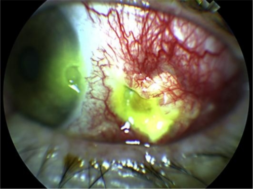
A corneal melt occurring in the context of an underlying collagen vascular disease is one of the clearest examples in ophthalmology of an eye finding that signals a life-threatening systemic problem, since progressive stromal thinning in this setting usually reflects active vasculitis, and treating the cornea alone without addressing the systemic disease is a well-documented path to both corneal perforation and, in severe cases, patient mortality.
Why Collagen Vascular Disease Causes Corneal Melting
The peripheral cornea is nourished in part by the limbal vascular arcade, and immune complex deposition and vasculitis affecting these vessels, along with local release of destructive enzymes including collagenases from inflammatory cells, leads to progressive breakdown of the corneal stroma.
This process, most classically seen as peripheral ulcerative keratitis, can also occur centrally and can progress rapidly from mild thinning to frank perforation if the underlying systemic inflammation is not controlled.
Associated Systemic Diseases
- Rheumatoid arthritis, the most common association, particularly in long-standing, seropositive, and erosive disease
- Granulomatosis with polyangiitis, in which corneal melt can be an early or presenting sign of active systemic vasculitis
- Relapsing polychondritis
- Systemic lupus erythematosus
- Polyarteritis nodosa and other systemic vasculitides
- Less commonly, inflammatory bowel disease and other autoimmune conditions
A corneal melt in a patient with known rheumatoid arthritis, particularly if the arthritis has been poorly controlled or has flared, should be treated as a marker of active systemic vasculitis until proven otherwise.
All-fit Slit-Lamp Adapter
Record and share exactly what you see at the slit lamp. One adapter fits any slit lamp or surgical microscope — and any smartphone.
From Choroida — the team behind this siteClinical Presentation
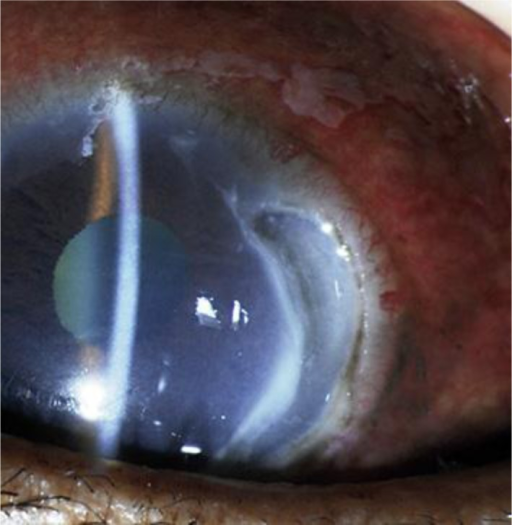
- Progressive thinning of the corneal stroma, often peripheral but occasionally central
- Pain, redness, and photophobia, though pain can be relatively mild given the severity of tissue destruction in some cases
- Crescentic guttering or ulceration at the limbus in peripheral ulcerative keratitis
- Risk of perforation if thinning progresses unchecked, sometimes over a period of days
- The sclera adjacent to the cornea may also show scleritis in the same eye, reflecting the shared vasculitic process
Differential Diagnosis
- Infectious keratitis, which must be excluded with culture, since immunosuppression is the treatment for the vasculitic process but would be harmful if an active infection were missed
- Mooren ulcer, a related but idiopathic form of peripheral ulcerative keratitis without an identified systemic association, requiring its own workup to confirm the absence of systemic disease before that diagnosis is accepted
- Neurotrophic keratopathy, which causes stromal thinning through a different mechanism, related to loss of corneal sensation rather than vasculitis
- Terrien marginal degeneration, a slowly progressive, typically painless and non-inflammatory peripheral thinning, distinguished by its different clinical course
Workup
- A full systemic vasculitis and autoimmune disease screen, mirroring the workup for scleritis: rheumatoid factor, anti-CCP antibodies, ANCA, ANA, complement levels, and inflammatory markers
- Corneal culture to exclude a superimposed or alternative infectious cause before starting systemic immunosuppression
- Coordination with rheumatology, both to confirm or characterize the underlying systemic disease and to manage systemic treatment
Management
Systemic Immunosuppression
This is the central and most important treatment, since corneal melt in this setting represents a manifestation of systemic vasculitis; treating the eye alone, without adequate systemic immunosuppression, has a well-documented history of poor outcomes, including recurrent melting and death from uncontrolled systemic vasculitis.
Systemic corticosteroids are used for initial control, followed by steroid-sparing agents such as cyclophosphamide, rituximab, or other immunosuppressants depending on the specific underlying disease, chosen and managed by rheumatology.
Local Ocular Measures
- Lubrication and, in severe cases, a bandage contact lens or tarsorrhaphy to protect a severely thinned cornea
- Topical collagenase inhibitors, such as topical medroxyprogesterone or doxycycline given systemically, sometimes used as adjuncts, though systemic immunosuppression remains the primary treatment
- Topical corticosteroids are used cautiously, since they can theoretically worsen stromal thinning if collagen synthesis is also being suppressed, and their use is balanced against the inflammatory component
Surgical Management
Impending or actual perforation may require tissue glue, an amniotic membrane patch, a corneal or scleral patch graft, or in severe cases a therapeutic penetrating keratoplasty, performed alongside, not instead of, systemic immunosuppression.
Prognosis
Ocular and systemic outcomes have improved substantially with prompt recognition and modern immunosuppressive therapy, but corneal melt in the setting of collagen vascular disease remains a marker of serious disease activity that requires urgent, coordinated ophthalmic and rheumatologic care.


Document what you see
Two smartphone imaging tools built for everyday clinic use — one for the slit lamp, one for the fundus.
From Choroida — the team behind this siteReferences
- Foster CS, Forstot SL, Wilson LA. Mortality rate in rheumatoid arthritis patients developing necrotizing scleritis or peripheral ulcerative keratitis. Ophthalmology. 1984;91:1253-1263.
- Messmer EM, Foster CS. Vasculitic peripheral ulcerative keratitis. Surv Ophthalmol. 1999;43:379-396.
- Tauber J, Sainz de la Maza M, Hoang-Xuan T, Foster CS. An analysis of therapeutic decision making regarding immunosuppressive chemotherapy for peripheral ulcerative keratitis. Cornea. 1990;9:66-73.
- Gomes JA, Rodrigues PC, Lopes LN. Mooren’s ulcer and infectious peripheral ulcerative keratitis. Semin Ophthalmol. 2014;29:327-337.
Test yourself
A few questions straight from this article.
-
Which process drives corneal stromal breakdown in collagen vascular disease?
Immune complex deposition and vasculitis affecting the limbal vascular arcade, together with destructive enzymes such as collagenases from inflammatory cells, progressively break down the corneal stroma. -
Which systemic disease is the most common association of corneal melt?
Rheumatoid arthritis is the most common association, particularly in long-standing, seropositive and erosive disease. -
Which corneal finding characterises peripheral ulcerative keratitis?
Peripheral ulcerative keratitis produces crescentic guttering or ulceration at the limbus as the stroma thins. -
What is true of the pain in corneal melt from collagen vascular disease?
Pain, redness and photophobia are typical, but in some cases the pain is relatively mild given the severity of the tissue destruction. -
Why is corneal culture taken before starting systemic immunosuppression for corneal melt?
Infectious keratitis must be excluded by culture, because immunosuppression treats the vasculitic process but would be harmful if an active infection were missed. -
How does Mooren ulcer differ from vasculitic corneal melt?
Mooren ulcer is a related but idiopathic form of peripheral ulcerative keratitis with no identified systemic association, and workup must confirm the absence of systemic disease before accepting it. -
Which feature points to Terrien marginal degeneration rather than vasculitic corneal melt?
Terrien marginal degeneration is a slowly progressive, typically painless and non-inflammatory peripheral thinning, distinguished by its different clinical course. -
What is the central treatment for corneal melt in collagen vascular disease?
Because the melt is a manifestation of systemic vasculitis, systemic immunosuppression is the central treatment; treating the eye alone has a documented history of recurrent melting and death from uncontrolled vasculitis. -
Which local measure helps protect a severely thinned cornea in this setting?
Local measures include lubrication and, in severe cases, a bandage contact lens or tarsorrhaphy to protect a severely thinned cornea. -
What does a corneal melt in collagen vascular disease signify about the patient overall?
Even with modern immunosuppressive therapy, corneal melt in collagen vascular disease remains a marker of serious disease activity requiring urgent, coordinated ophthalmic and rheumatologic care.

Writes and reviews the clinical articles on Educate. Every post is checked against peer-reviewed sources before it goes live.


