
A child whose eye turns inward is often brought in because of how it looks. The reason to act is what it is doing to their vision.
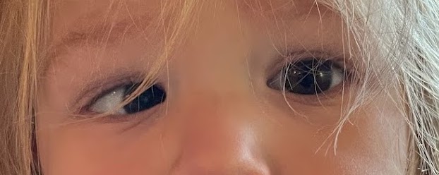
Esotropia is inward deviation of one eye, and it is the most common form of strabismus in childhood.
A misaligned eye in a developing visual system produces two problems at once: the brain suppresses the deviating eye, and binocular vision fails to develop.
One form of esotropia disappears entirely with a pair of glasses, because the deviation is driven by uncorrected hypermetropia.
Another form, appearing suddenly in an older child or adult, can be the first sign of intracranial disease.
Separating these types on examination is what determines whether the answer is spectacles, surgery, or a scan.
What Is Esotropia?
Esotropia is a manifest convergent misalignment in which one eye deviates nasally while the other fixates.
The clinically important types are:
- Infantile (congenital) esotropia – onset before six months, with a large, stable angle and generally low hypermetropia
- Accommodative esotropia – onset typically between two and four years, driven by uncorrected hypermetropia, and improving or fully correcting with glasses
- Partially accommodative esotropia – the angle reduces with glasses but a residual deviation remains
- Sensory esotropia – secondary to poor vision in one eye from cataract, corneal opacity or retinal disease
- Acute acquired comitant esotropia – sudden onset with diplopia, which requires exclusion of intracranial pathology
- Incomitant esotropia from a sixth nerve palsy – the angle varies with gaze direction and abduction is limited
The single most useful distinction on examination is comitant versus incomitant, because an incomitant deviation points to a nerve palsy or restriction rather than a developmental squint.
Epidemiology
Esotropia is the predominant strabismus pattern in childhood in most populations.
- Strabismus affects a small but significant percentage of children, and esotropia accounts for the majority of cases in many series
- Accommodative esotropia is the commonest type overall and typically presents in the preschool years
- Infantile esotropia presents by definition within the first six months of life
- A family history of strabismus or significant refractive error is common
- Prematurity, neurological impairment and developmental delay all increase risk
Because amblyopia risk falls sharply with age at treatment, the practical value of early detection in this condition is very high.
All-fit Slit-Lamp Adapter
Record and share exactly what you see at the slit lamp. One adapter fits any slit lamp or surgical microscope — and any smartphone.
From Choroida — the team behind this sitePathophysiology
The mechanisms differ substantially between types, which is why they are managed so differently.
- In accommodative esotropia, uncorrected hypermetropia forces the child to accommodate to see clearly; accommodation is linked to convergence, and the excess convergence produces the inward deviation
- A high AC/A ratio – excess convergence per unit of accommodation – explains cases where the deviation is much larger at near than at distance
- In infantile esotropia, the deviation reflects a primary failure of normal binocular development rather than a refractive drive
- In sensory esotropia, one eye loses the visual input needed to maintain alignment and drifts
- Once an eye is deviated during visual immaturity, the brain suppresses its image to avoid diplopia, producing strabismic amblyopia and loss of stereopsis
The accommodative mechanism is the reason a full cycloplegic refraction is mandatory in every child with esotropia – missing hypermetropia means missing a deviation that glasses alone would cure.
Risk Factors
Ocular and Refractive
- Significant uncorrected hypermetropia
- Anisometropia
- Any cause of unilateral visual loss – cataract, corneal scar, retinal or optic nerve disease
Systemic and Perinatal
- Prematurity and low birth weight
- Cerebral palsy, developmental delay and other neurological conditions
- Family history of strabismus or amblyopia
- Genetic syndromes, notably trisomy 21
Any child presenting with esotropia needs a dilated fundus examination, because a sensory esotropia can be the first presenting sign of retinoblastoma.
Clinical Presentation
Symptoms
- A visible inward turn of one eye, often intermittent at first and more obvious when the child is tired or unwell
- Head turn toward the affected side in a sixth nerve palsy, adopted to maintain single vision
- Diplopia in older children and adults, but characteristically absent in young children because of suppression
- Eye rubbing, closing one eye in bright light, or a parental report that the eyes “do not work together”
Examination Findings
- An asymmetric corneal light reflex, displaced temporally in the deviating eye (Hirschberg test)
- A cover test demonstrating outward refixation movement of the deviated eye when the fixing eye is covered
- Measurement of the angle at distance and near using prism cover testing
- Assessment of ocular motility for limitation of abduction, which indicates a sixth nerve palsy rather than a comitant squint
- Cycloplegic refraction, which is essential and frequently reveals significant hypermetropia
- Dilated fundus examination to exclude an organic cause of sensory esotropia
Prominent epicanthal folds and a broad nasal bridge can mimic esotropia convincingly – pseudoesotropia is distinguished by a symmetric corneal light reflex and a normal cover test.
Diagnostic Evaluation
Orthoptic Assessment
- Visual acuity measured with age-appropriate tests, comparing the two eyes for evidence of amblyopia
- Prism cover test at distance and near to quantify the deviation and identify a high AC/A ratio
- Assessment of stereopsis and binocular function
Refraction
- Cycloplegic refraction in every case – non-cycloplegic refraction in a child will systematically underestimate hypermetropia
- Repeat refraction over time, since refractive status changes as the child grows
When Neuroimaging Is Required
- Acute acquired comitant esotropia with diplopia, particularly in an older child
- Any incomitant deviation with limited abduction suggesting a sixth nerve palsy
- Associated neurological signs, headache, papilledema or nystagmus
A sixth nerve palsy is a false localising sign of raised intracranial pressure, so an abduction deficit should always prompt a look at the optic discs.
Differential Diagnosis
Conditions that may be confused with esotropia include:
- Pseudoesotropia from epicanthal folds – normal corneal reflex and no movement on cover testing
- Sixth nerve palsy – incomitant, with limited abduction
- Duane retraction syndrome – globe retraction and palpebral fissure narrowing on adduction
- Mobius syndrome and other congenital cranial dysinnervation disorders
- Thyroid eye disease with medial rectus restriction, typically in adults
- Convergence spasm – variable, with associated miosis and accommodative changes
Distinguishing pseudoesotropia matters practically, because it is common, entirely benign, and needs only explanation rather than treatment.
Management
Optical Correction
- Full hypermetropic correction based on cycloplegic refraction is first-line, and fully corrects the deviation in true accommodative esotropia
- Bifocals may be used where the deviation is significantly larger at near due to a high AC/A ratio
- Glasses should be given before considering surgery in any accommodative component
Amblyopia Treatment
- Patching or atropine penalisation of the better-seeing eye to treat strabismic amblyopia
- Amblyopia should be treated before surgical alignment, since a stable visual outcome depends on it
- Treatment is most effective when started young and continued consistently
Surgery and Other Options
- Strabismus surgery – typically bilateral medial rectus recession – for infantile esotropia and for residual non-accommodative deviation
- Early surgery in infantile esotropia aims to give the best chance of some binocular function
- Botulinum toxin injection has a role in selected cases, including some acute and post-palsy deviations
- Treatment of the underlying cause in sensory and paralytic esotropia
The order matters: refraction first, then amblyopia, then surgery for whatever deviation remains – operating on an uncorrected accommodative component risks overcorrection.
Prognosis
Outcomes are good when treatment starts early and is followed through.
- Accommodative esotropia treated with appropriate glasses has an excellent prognosis, though spectacle dependence often continues for years
- Amblyopia responds well to treatment in young children, and progressively less well with increasing age
- Infantile esotropia can usually be aligned surgically, but high-grade stereopsis is rarely achieved
- Recurrence or a change in the deviation over time is common, and repeat surgery is sometimes needed
- Sensory esotropia carries the prognosis of the underlying ocular disease
Alignment and vision are separate goals – a cosmetically straight eye that was never treated for amblyopia is still a functionally poor result.


Document what you see
Two smartphone imaging tools built for everyday clinic use — one for the slit lamp, one for the fundus.
From Choroida — the team behind this siteReferences
- Pediatric Eye Disease Investigator Group. A randomized trial of atropine vs patching for treatment of moderate amblyopia in children. Archives of Ophthalmology. 2002.
- American Academy of Ophthalmology. Esotropia and Exotropia Preferred Practice Pattern.
- Birch EE. Amblyopia and binocular vision. Progress in Retinal and Eye Research. 2013.
- Esotropia. StatPearls, NCBI Bookshelf.
- Esotropia. EyeWiki, American Academy of Ophthalmology.
Test yourself
A few questions straight from this article.
-
How is esotropia defined?
Esotropia is a manifest convergent misalignment in which one eye deviates nasally while the fellow eye fixates, and it is the commonest childhood strabismus. -
By what age does infantile esotropia present by definition?
Infantile esotropia is defined by onset before six months, typically with a large, stable angle and generally low hypermetropia. -
What mechanism produces the inward deviation in accommodative esotropia?
Uncorrected hypermetropia forces the child to accommodate; because accommodation is linked to convergence, the excess convergence produces the inward deviation. -
A high AC/A ratio in esotropia produces which pattern on prism cover testing?
A high AC/A ratio means excess convergence per unit of accommodation, so the deviation is much larger at near than at distance and may need bifocals. -
What does the Hirschberg corneal light reflex show in an eye with esotropia?
Because the eye is turned nasally, the corneal light reflex sits temporally in the deviating eye, giving the asymmetric reflex of the Hirschberg test. -
How is pseudoesotropia from epicanthal folds distinguished from a true esotropia?
Prominent epicanthal folds and a broad nasal bridge mimic esotropia convincingly, but the corneal reflex stays symmetric and the cover test shows no refixation movement. -
In a child with esotropia and limited abduction, why must the optic discs be examined?
An abduction deficit points to a sixth nerve palsy, which is a false localising sign of raised intracranial pressure, so the optic discs should always be inspected. -
Why does every child presenting with esotropia need a dilated fundus examination?
Sensory esotropia follows poor vision in one eye, and retinoblastoma is among the organic causes that must be excluded on dilated fundus examination. -
What is the first-line treatment for a child with accommodative esotropia?
Full hypermetropic spectacles fully correct a true accommodative esotropia, and operating on an uncorrected accommodative component risks overcorrection. -
What is the usual outcome after surgery for infantile esotropia?
Infantile esotropia can usually be aligned surgically, but high-grade stereopsis is rarely achieved and recurrence or drift over time is common.

Writes and reviews the clinical articles on Educate. Every post is checked against peer-reviewed sources before it goes live.


