
Senior-Loken syndrome combines nephronophthisis, a progressive tubulointerstitial kidney disease, with a retinal dystrophy, and both organ systems fail for the same underlying reason: a defect in the primary cilium, a structure essential to both renal tubular cells and retinal photoreceptors.
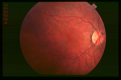
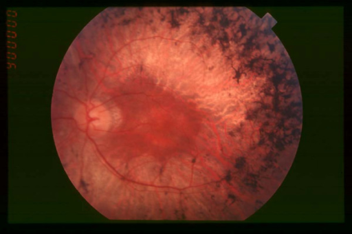
Recognizing the retinal findings in a child with unexplained kidney disease, or vice versa, can bring forward the diagnosis before end-stage renal disease develops silently.
A Shared Ciliopathy
The connecting cilium of the photoreceptor and the primary cilium of the renal tubular cell share overlapping molecular machinery, and mutations in genes that disrupt ciliary function, including several NPHP genes, cause dysfunction in both tissues simultaneously.
This is why Senior-Loken syndrome is grouped with other ciliopathies, a family of conditions that also includes Bardet-Biedl syndrome and Joubert syndrome, each affecting an overlapping but distinct set of organs depending on which ciliary gene and function is disrupted.
Renal Features
Nephronophthisis is a chronic tubulointerstitial nephropathy that progresses to end-stage renal disease, typically in childhood or adolescence for the more common juvenile form, though infantile and adolescent forms with different ages of onset also occur.
Early signs are often subtle: polyuria, polydipsia, and growth impairment from impaired urinary concentrating ability, which can be missed until renal function has already declined significantly, since overt proteinuria and hypertension are characteristically late or absent findings compared with most other causes of chronic kidney disease.
Ocular Features
The retinal dystrophy in Senior-Loken syndrome ranges in severity and age of onset.
- A Leber congenital amaurosis-like presentation, with severe visual impairment and nystagmus from infancy
- A later-onset retinitis pigmentosa-like pattern, with progressive night blindness and peripheral field loss beginning in later childhood
- Electroretinography shows severely reduced or nonrecordable responses, consistent with a rod-cone or generalized photoreceptor dystrophy
- Fundus findings can range from a near-normal appearance early on to bone-spicule pigmentation and vascular attenuation as the disease progresses
The severity and pattern of retinal involvement do not always correlate closely with the severity of renal disease, so normal-looking kidneys do not guarantee mild retinal disease or vice versa.
Diagnosis
The combination of an unexplained retinal dystrophy with renal concentrating defects, growth impairment, or unexplained chronic kidney disease should prompt consideration of Senior-Loken syndrome.
- Renal ultrasound, which may show small, echogenic kidneys with cysts, though this can be a late finding
- Formal renal function testing, including urine concentrating ability
- Electroretinography and OCT to characterize the retinal dystrophy
- Genetic testing for NPHP and related ciliopathy genes, which confirms the diagnosis and can guide family counseling
Because the renal disease can progress silently, children diagnosed with an early-onset retinal dystrophy of unclear cause should have renal function screened even in the absence of urinary symptoms.
Management
Renal Disease
Management follows standard nephrology care for chronic kidney disease, with dialysis and eventual renal transplantation when end-stage renal disease develops.
Regular monitoring of growth, blood pressure, and renal function allows earlier intervention before uremic complications develop.
Ocular Disease
There is no treatment that restores photoreceptor function, so ocular management focuses on low-vision support, correction of refractive error, and genetic counseling.
Gene-specific therapies for inherited retinal disease are an active area of research, and some ciliopathy-related retinal genes are being studied as targets, which makes accurate genetic diagnosis potentially relevant to future treatment eligibility.
Prognosis
Renal disease typically progresses to end-stage renal failure requiring transplantation, usually by adolescence or young adulthood for the more common juvenile form.
Visual prognosis depends on the specific retinal phenotype, ranging from severe early childhood visual impairment to a more gradual decline extending into adulthood.


Document what you see
Two smartphone imaging tools built for everyday clinic use — one for the slit lamp, one for the fundus.
From Choroida — the team behind this siteReferences
- Senior B, Friedmann AI, Braudo JL. Juvenile familial nephropathy with tapetoretinal degeneration: a new oculorenal dystrophy. Am J Ophthalmol. 1961;52:625-633.
- Loken AC, Hanssen O, Halvorsen S, Jolster NJ. Hereditary renal dysplasia and blindness. Acta Paediatr. 1961;50:177-184.
- Hildebrandt F, Zhou W. Nephronophthisis-associated ciliopathies. J Am Soc Nephrol. 2007;18:1855-1871.
- Otto EA, Loeys B, Khanna H, et al. Nephrocystin-5, a ciliary IQ domain protein, is mutated in Senior-Loken syndrome and interacts with RPGR and calmodulin. Nat Genet. 2005;37:282-288.
