
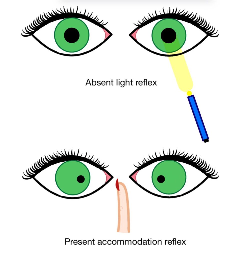
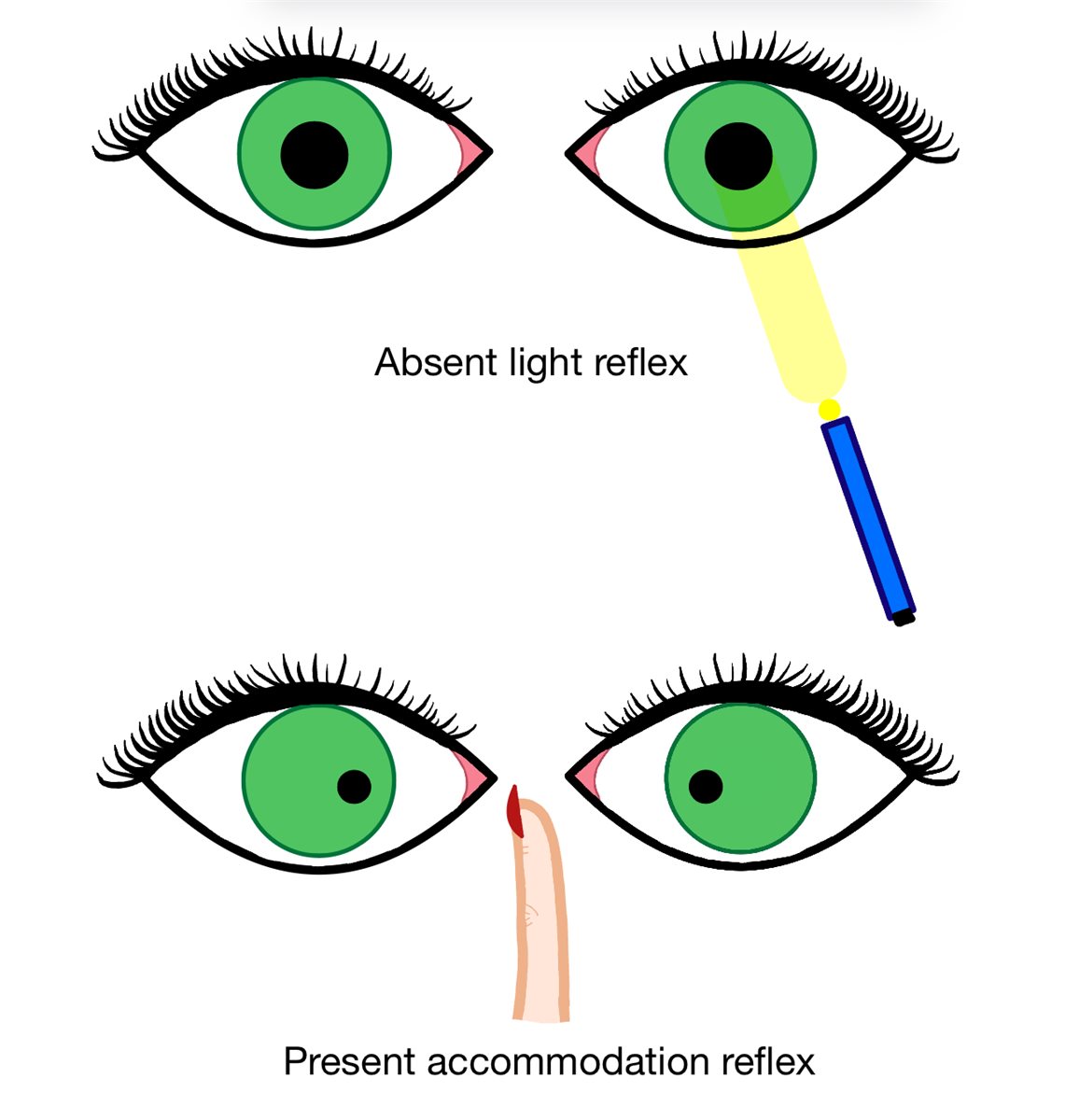
An Argyll Robertson pupil is a small, irregular pupil that fails to constrict to light but constricts normally when the patient looks at a near object, a dissociation between two reflex pathways that once made this sign one of the most reliable bedside indicators of neurosyphilis.
Genuine light-near dissociation of this specific type is now rare in everyday practice, largely because tertiary syphilis itself has become far less common, but the underlying reflex pathways it tests remain clinically important, and recognising the pattern still matters whenever it does appear.
The Reflex Pathways Involved
The pupillary light reflex and the near (accommodative) reflex both ultimately act on the same pupillary sphincter muscle, but they are driven by anatomically distinct pathways within the midbrain before converging on the final common pathway through the third cranial nerve.
A lesion affecting the light reflex pathway selectively, most classically at the level of the pretectal nucleus, while sparing the separate pathway mediating the near response, produces light-near dissociation: no pupillary constriction to a light stimulus, but normal constriction on convergence for near fixation.
In neurosyphilis, this selective damage is attributed to chronic inflammatory change affecting the pretectal region, though the precise mechanism has never been fully established with certainty.
Clinical Features
- Bilateral involvement is typical, though the two pupils are often asymmetric in size and shape rather than identically affected
- Small, irregular pupils, sometimes nicknamed “prostitute’s pupils” in older texts because they “accommodate but do not react,” a crude but genuinely memorable way to recall the light-near dissociation
- Poor pupillary dilation in the dark is common, related to the same underlying structural change
- Absent or minimal response to light, whether direct or consensual
- Normal, brisk constriction with near fixation and convergence, the defining dissociation
All-fit Slit-Lamp Adapter
Record and share exactly what you see at the slit lamp. One adapter fits any slit lamp or surgical microscope — and any smartphone.
From Choroida — the team behind this siteDifferential Diagnosis of Light-Near Dissociation
- Adie tonic pupil, typically unilateral, initially larger rather than smaller than the fellow eye, with a slow, tonic near response and denervation supersensitivity to dilute pilocarpine, a very different clinical and pharmacological profile from Argyll Robertson pupils
- Dorsal midbrain (Parinaud) syndrome, associated with additional signs including upgaze palsy and convergence-retraction nystagmus, reflecting a lesion at the level of the dorsal midbrain rather than a purely pupillary process
- Severe bilateral afferent visual pathway disease, which can produce an apparent light-near dissociation simply because the light reflex pathway is impaired by profound vision loss rather than by a lesion specific to the pupillary reflex arc itself
- Diabetic autonomic neuropathy, occasionally producing a similar dissociation through a different underlying mechanism
Distinguishing Adie tonic pupil from a true Argyll Robertson pupil is the most clinically important discrimination on this list, since the two point toward entirely different underlying diseases and require different investigation pathways.
Diagnostic Evaluation
Serological testing for syphilis, including both a non-treponemal test such as RPR or VDRL and a confirmatory treponemal-specific test, is the essential first step whenever a true Argyll Robertson pupil is suspected.
Cerebrospinal fluid examination, including CSF VDRL, is indicated when neurosyphilis is suspected, both to confirm central nervous system involvement and to guide treatment duration and monitoring of response.
A broader neurological assessment for other signs of tabes dorsalis or general paresis, the classic tertiary neurosyphilis syndromes historically associated with this pupillary finding, is appropriate once the diagnosis is confirmed.
Management
Treatment addresses the underlying neurosyphilis, typically with high-dose intravenous penicillin, following current sexually transmitted infection treatment guidelines and coordinated with infectious disease specialists.
The pupillary abnormality itself does not require any specific ocular treatment and, importantly, does not reliably resolve even with successful treatment of the underlying infection, since it reflects structural damage to the pretectal pathway rather than an active, reversible inflammatory process.
Ongoing monitoring for other manifestations of tertiary syphilis and appropriate follow-up serology to confirm adequate treatment response are part of standard management, coordinated with the team managing the systemic infection.
Prognosis
The pupillary finding itself causes minimal functional visual impairment, since visual acuity and the near response are preserved. Patients occasionally notice mild glare or light sensitivity related to the poor pupillary light response, but this is rarely a significant complaint.
The pupillary abnormality is generally permanent even after adequate treatment of the underlying neurosyphilis, reflecting fixed structural damage rather than ongoing active disease, and this permanence should be part of the counselling given once the diagnosis and treatment plan are established.
Overall prognosis is determined by the extent of the underlying neurosyphilis and how promptly it is treated, since untreated tertiary syphilis carries the risk of progressive and serious neurological disease well beyond the pupillary finding that first brought it to attention.


Document what you see
Two smartphone imaging tools built for everyday clinic use — one for the slit lamp, one for the fundus.
From Choroida — the team behind this siteReferences
- Loewenfeld IE. The Pupil: Anatomy, Physiology, and Clinical Applications. Butterworth-Heinemann.
- Thompson HS, Kardon RH. The Argyll Robertson pupil. Journal of Neuro-Ophthalmology. 2006.
- Ropper AH, Samuels MA, Klein JP. Adams and Victor’s Principles of Neurology. McGraw-Hill.
- Argyll Robertson Pupil. EyeWiki, American Academy of Ophthalmology.
- Neurosyphilis. StatPearls, NCBI Bookshelf.
Test yourself
A few questions straight from this article.
-
Which pupillary pattern defines an Argyll Robertson pupil?
The Argyll Robertson pupil is small and irregular, shows absent or minimal response to light, yet constricts briskly on convergence for near fixation. -
Where is the lesion classically located in Argyll Robertson pupil?
Selective damage at the pretectal level interrupts the light reflex pathway while sparing the separate near-response pathway, producing light-near dissociation. -
What is the typical laterality of Argyll Robertson pupils?
Bilateral involvement is typical, though the two pupils commonly differ in size and shape rather than being identically affected. -
How do Argyll Robertson pupils behave in dim illumination?
Poor dilation in the dark is common and reflects the same underlying structural change that abolishes the light response. -
Which feature distinguishes an Adie tonic pupil from an Argyll Robertson pupil?
Adie tonic pupil is usually unilateral, initially larger than the fellow pupil, has a slow tonic near response and shows denervation supersensitivity to dilute pilocarpine. -
Which additional signs point to dorsal midbrain syndrome rather than Argyll Robertson pupil?
Dorsal midbrain (Parinaud) syndrome adds upgaze palsy and convergence-retraction nystagmus, reflecting a dorsal midbrain lesion rather than a purely pupillary process. -
What is the essential first investigation when a true Argyll Robertson pupil is suspected?
Serology combining a non-treponemal test such as RPR or VDRL with a confirmatory treponemal-specific test is the essential first step. -
What does cerebrospinal fluid VDRL testing add when neurosyphilis is suspected?
CSF examination including CSF VDRL confirms central nervous system involvement and guides treatment duration and monitoring of response. -
How is the neurosyphilis underlying an Argyll Robertson pupil treated?
Treatment targets the underlying neurosyphilis with high-dose intravenous penicillin, following current guidelines and coordinated with infectious disease specialists. -
What happens to an Argyll Robertson pupil after successful treatment of neurosyphilis?
The pupillary abnormality reflects fixed structural damage to the pretectal pathway rather than active inflammation, so it does not reliably resolve with treatment.

Writes and reviews the clinical articles on Educate. Every post is checked against peer-reviewed sources before it goes live.


