
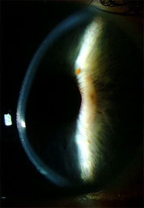
Post-LASIK ectasia is progressive corneal thinning and steepening occurring after refractive surgery, discussed more generally in relation to LASIK candidate selection in its own dedicated article on this site, and it represents the single complication that preoperative screening is most specifically designed to prevent.
The condition looks and behaves much like keratoconus, discussed in its own dedicated article on this site, but occurs in a cornea that has already been surgically thinned and structurally weakened by the creation of a LASIK flap and the removal of stromal tissue during the original procedure.
Understanding why certain corneas are vulnerable to this complication, and how screening has evolved specifically to identify them beforehand, is central to understanding both the risk and the now much-reduced incidence of ectasia in modern refractive surgery practice.
Radial keratotomy, discussed in its own dedicated article on this site, causes a different, older form of iatrogenic corneal weakening, and while the two conditions are not identical, both illustrate the same broader principle that refractive corneal surgery permanently alters the cornea’s biomechanics in ways that can have long-term consequences.
Mechanism
LASIK removes stromal tissue to reshape the cornea and additionally creates a flap that, while it heals, never regains the full biomechanical strength of unwounded corneal tissue, meaning every LASIK-treated cornea is somewhat structurally weaker than it was before surgery.
In most corneas, this reduction in strength is well tolerated, since there is ample residual stromal tissue to maintain adequate structural integrity after the treatment and flap creation.
In a cornea that was already biomechanically weaker than average before surgery, whether from unrecognized subclinical keratoconus or simply inherently thinner or less rigid tissue, this combination of stromal removal and flap creation can tip the balance toward progressive, ectasia-like thinning and steepening over the months to years following surgery.
Risk Factors
- Preoperative corneal topography or tomography showing subtle abnormalities suggestive of forme fruste, or subclinical, keratoconus, the single most important risk factor to identify before surgery
- Insufficient residual stromal bed thickness after the planned treatment, generally requiring more tissue removal relative to the cornea’s total thickness than is considered safe
- Younger patient age at the time of surgery, associated with a somewhat higher reported risk in some series
- High preoperative myopia, requiring deeper tissue ablation to achieve the intended refractive correction and therefore leaving comparatively less residual stromal support
- A family history of keratoconus, raising concern for an undetected genetic predisposition even when preoperative topography appears reassuring
All-fit Slit-Lamp Adapter
Record and share exactly what you see at the slit lamp. One adapter fits any slit lamp or surgical microscope — and any smartphone.
From Choroida — the team behind this siteWhy Preoperative Screening Matters So Much
Corneal topography and, increasingly, more detailed tomography mapping both the anterior and posterior corneal surfaces are used specifically to detect the subtle irregularities characteristic of subclinical keratoconus before it becomes clinically obvious.
Calculating and respecting a minimum residual stromal bed thickness after the planned treatment is a standard, deliberate part of surgical planning, directly aimed at avoiding the specific combination of risk factors known to predispose to ectasia.
Modern refractive surgery screening protocols, incorporating these more sophisticated imaging and calculation methods, have meaningfully reduced the incidence of post-LASIK ectasia compared to the earlier years of the procedure’s widespread use, when screening tools were considerably less refined.
Even with careful screening, a small residual risk remains, since current tools cannot identify every biomechanically vulnerable cornea with complete certainty, which is an honest point worth including in preoperative counseling rather than presenting screening as an absolute guarantee.
Clinical Presentation
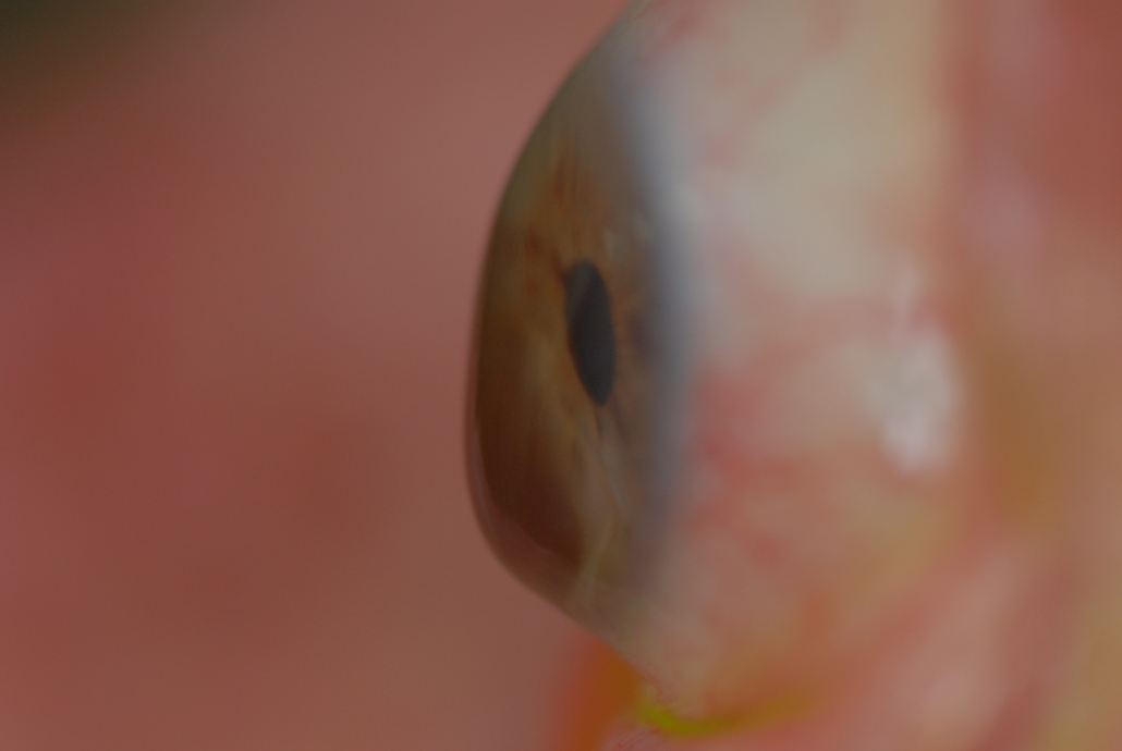
Progressive myopic or astigmatic shift in the months to years after an initially successful LASIK procedure is the hallmark presenting feature, often accompanied by decreasing best-corrected visual acuity as the cornea’s irregular astigmatism increases.
Topographic findings mirror those of keratoconus, including progressive inferior or central steepening and thinning, and comparison with the patient’s own preoperative topography, when available, is invaluable for confirming genuine progression rather than an incidental finding.
Symptoms can develop gradually enough that they are sometimes initially attributed to a simple, expected refractive regression rather than recognized as a progressive structural process requiring its own specific management.
Management
Corneal collagen cross-linking, discussed in its own dedicated article on this site, is used to halt progression in post-LASIK ectasia, following the same general principle applied to primary keratoconus.
Rigid gas-permeable or scleral contact lenses, discussed in relation to scleral lens fitting in its own dedicated article on this site, address the irregular astigmatism that glasses cannot adequately correct once significant ectasia has developed.
Corneal transplantation, whether penetrating or lamellar, both discussed in their own dedicated articles on this site, is reserved for advanced cases where contact lenses can no longer provide functional vision and cross-linking has not adequately halted progression.
Early detection through diligent postoperative follow-up remains valuable even after screening, since catching progression early gives cross-linking the best chance of halting the process before it advances to the point of needing contact lenses or transplantation.


Document what you see
Two smartphone imaging tools built for everyday clinic use — one for the slit lamp, one for the fundus.
From Choroida — the team behind this siteReferences
- Randleman JB, Woodward M, Lynn MJ, Stulting RD. Risk assessment for ectasia after corneal refractive surgery. Ophthalmology.
- American Academy of Ophthalmology. Basic and Clinical Science Course, Section 13: Refractive Surgery.
- Binder PS. Analysis of ectasia after laser in situ keratomileusis: risk factors. Journal of Cataract and Refractive Surgery.
Test yourself
A few questions straight from this article.
-
Post-LASIK ectasia is characterised by which corneal change after refractive surgery?
Post-LASIK ectasia is progressive corneal thinning and steepening after refractive surgery, behaving much like keratoconus but in a cornea already surgically thinned and weakened. -
Which two elements of LASIK combine to weaken the cornea biomechanically?
LASIK removes stromal tissue and creates a flap that, although it heals, never regains the full biomechanical strength of unwounded cornea, so every treated cornea is somewhat weaker. -
Which preoperative finding is the single most important risk factor for post-LASIK ectasia?
Subtle topographic or tomographic abnormalities suggesting forme fruste, or subclinical, keratoconus are the single most important risk factor to identify before refractive surgery. -
Which surgical planning error predisposes a LASIK patient to corneal ectasia?
Insufficient residual stromal bed thickness after the planned treatment is a recognised risk factor, which is why calculating and respecting a minimum residual bed is standard planning. -
Why does high preoperative myopia increase the risk of post-LASIK ectasia?
High myopia requires deeper tissue ablation to achieve the intended correction, leaving comparatively less residual stromal support behind. -
What does corneal tomography add over topography when screening for ectasia risk?
Increasingly detailed tomography maps both the anterior and posterior corneal surfaces to detect the subtle irregularities of subclinical keratoconus before it becomes clinically obvious. -
When is corneal transplantation considered in post-LASIK ectasia?
Penetrating or lamellar transplantation is reserved for advanced cases where contact lenses can no longer provide functional vision and cross-linking has not halted progression. -
What is the hallmark presenting feature of post-LASIK ectasia?
A progressive myopic or astigmatic shift in the months to years after an initially successful LASIK, often with falling best-corrected acuity, is the hallmark presentation. -
Gradually developing post-LASIK ectasia is sometimes initially mistaken for which benign process?
Symptoms can develop gradually enough that they are attributed to expected refractive regression rather than recognised as a progressive structural process needing specific management. -
Which treatment is used to halt progression of post-LASIK ectasia?
Corneal collagen cross-linking halts progression in post-LASIK ectasia, following the same principle applied in primary keratoconus, and works best when progression is caught early.

Writes and reviews the clinical articles on Educate. Every post is checked against peer-reviewed sources before it goes live.


