
Pseudophakic retinal detachment is a rhegmatogenous detachment in an eye that has had cataract surgery, and it is one of the most important late complications of an otherwise successful operation.
The absolute risk is low, below one percent in most large series, but the number of cataract operations is so large that pseudophakic detachments make up a significant share of all detachments.
The breaks are often small and anterior, the view is poor because of capsular and lens edge reflections, and the detachment is frequently more advanced by the time the patient is seen.
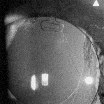
What Is Pseudophakic Retinal Detachment?
The retinal detachment develops through the same mechanism as other rhegmatogenous detachments, with a full-thickness break and liquefied vitreous.
Cataract surgery changes the vitreous, and posterior vitreous detachment occurs earlier and more frequently after surgery.
The detachment can appear weeks to years after surgery, and the risk continues over many years.
Risk Factors
Large database studies and national registries identify several factors.
- Posterior capsule rupture with vitreous loss, which raises the risk many times over
- Male sex
- Younger age at surgery, particularly under 65
- Longer axial length and high myopia
- Previous retinal detachment in the fellow eye
- Lattice degeneration and known retinal tears
- Nd:YAG capsulotomy, which has been associated with a higher risk of detachment in several studies (see YAG laser capsulotomy)
- Zonular weakness and complicated surgery
- Family history of detachment
A large population study using the nonoperated fellow eye as a control found increased risk in operated eyes, particularly in the first years after surgery.
Clear lens extraction in highly myopic eyes carries a higher risk of detachment than standard cataract surgery.
Fundus Explorer Pro
Photograph the retinal findings described here with the phone already in your pocket — 22 D optics and built-in illumination in one handheld unit.
From Choroida — the team behind this siteClinical Features
Symptoms are the same as in phakic detachment: new floaters, flashes, and a shadow that spreads across the field.
A few points are specific to pseudophakic eyes.
- The breaks are often small, multiple, and located anteriorly near the ora serrata or the vitreous base
- The detached retina is often bullous with fewer signs of chronicity
- Posterior capsule opacification, the IOL edge, and anterior capsular rim can obscure the peripheral view
- Pigment cells in the anterior vitreous are a valuable clue
- Vitreous hemorrhage and inflammation may hide the retina
- PVR develops earlier in some eyes
Patients who have had cataract surgery may attribute new floaters to the surgery and delay presenting, so counseling before and after surgery matters.
Examination
A high index of suspicion and careful technique are required.
- Full dilation with scleral depression and indirect ophthalmoscopy, checking the periphery through the pupil with the lens edge and capsule in mind
- Slit-lamp examination with a fundus contact lens or 90 D lens for the anterior peripheral retina
- Examination of the fellow eye, since bilateral disease is common
- B-scan ultrasonography when the view is poor
- Widefield imaging to document the extent
A small break may be visible only when the eye is turned and the depressor is applied over the periphery, so examination should be systematic and clock-hour based.
Management
Surgical Options
- Pars plana vitrectomy is the most commonly chosen operation, since it allows removal of vitreous and inspection of the periphery through a clear view (see pars plana vitrectomy)
- Scleral buckling can be used alone or combined with vitrectomy, and it supports the vitreous base and peripheral breaks (see scleral buckle surgery)
- Pneumatic retinopexy is possible for selected eyes with a clearly visible single break in the superior retina, although failure is more likely when breaks are multiple or hard to find
The choice depends on break location, media clarity, PVR, surgeon preference, and the patient’s ability to position.
The PIVOT trial included pseudophakic eyes, and single-surgery success favored vitrectomy over pneumatic retinopexy, while acuity and metamorphopsia outcomes favored pneumatic retinopexy.
Perioperative Points
- Look for retained lens material and posterior capsule tears, which may contribute to breaks
- Treat anterior breaks and the vitreous base carefully, since missed breaks cause failure
- Consider combined buckling when breaks are inferior or numerous, or when PVR is present
- Choose tamponade according to break location and the patient’s ability to position
Outcomes
Single-surgery success is high in most series, but redetachment from PVR and missed breaks is more common than in some other detachments.
Visual outcome depends on the status of the macula and the duration of detachment.
Common Clinical Scenarios
- A patient with new floaters two years after uncomplicated surgery, and a hazy capsule: examine the periphery carefully, since anterior breaks are easily missed
- A patient who had posterior capsule rupture and now reports flashes: the risk is high, and examination should be prompt and thorough
- A patient with detachment in one eye and cataract surgery planned in the other: consider a peripheral retinal assessment first, and discuss the risks
- A patient with detachment after capsulotomy: search for breaks at the vitreous base, and examine the fellow eye
In each of these cases the earlier the retinal specialist sees the patient, the better the anatomic and visual result.
Prevention and Counseling
Patients should be told before cataract surgery that there is a small risk of detachment, and after surgery they should know what to look for.
- New flashes, floaters, or shadow need same-day examination
- Highly myopic patients and those with intraoperative complications need a peripheral retinal examination
- Discuss the risks and benefits of YAG capsulotomy in patients with risk factors
- Evaluate the fellow eye when a detachment is found
- Treat symptomatic breaks promptly (see laser retinopexy)


Document what you see
Two smartphone imaging tools built for everyday clinic use — one for the slit lamp, one for the fundus.
From Choroida — the team behind this siteReferences
- Clark A, Morlet N, Ng JQ, Preen DB, Semmens JB. Risk for retinal detachment after phacoemulsification: a whole-population study of cataract surgery outcomes. Arch Ophthalmol. 2012;130:882-888.
- Bjerrum SS, Mikkelsen KL, La Cour M. Risk of pseudophakic retinal detachment in 202,226 patients using the fellow nonoperated eye as reference. Ophthalmology. 2013;120:2573-2579.
- Daien V, Le Pape A, Heve D, Carriere I, Villain M. Incidence, risk factors, and impact of age on retinal detachment after cataract surgery in France: a national population study. Ophthalmology. 2015;122:2179-2185.
- Powe NR, Schein OD, Gieser SC, et al. Synthesis of the literature on visual acuity and complications following cataract extraction with intraocular lens implantation. Arch Ophthalmol. 1994;112:239-252.
- Hillier RJ, Felfeli T, Berger AR, et al. The Pneumatic Retinopexy versus Vitrectomy for the Management of Primary Rhegmatogenous Retinal Detachment Outcomes Randomized Trial (PIVOT). Ophthalmology. 2019;126:531-539.
- Kuhn F, Aylward B. Rhegmatogenous retinal detachment: a reappraisal of its pathophysiology and treatment. Ophthalmic Res. 2014;51:15-31.
