
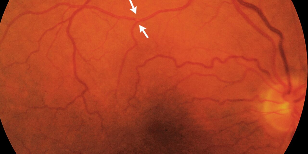
Amaurosis fugax is a transient loss of vision in one eye that lasts seconds to minutes and then resolves completely.
The most important cause is embolism or hypoperfusion from carotid or cardiac disease, and in that setting the episode is a retinal transient ischemic attack that carries a risk of stroke.
Giant cell arteritis is the other diagnosis that cannot be missed, because untreated it can cause permanent blindness within days.
Many other conditions produce brief monocular visual loss, so the history decides how urgent the workup is.
What Is Amaurosis Fugax?
The term describes a symptom and not a disease.
Patients typically say that a curtain or shade descended over the vision of one eye, or that vision dimmed or blurred, and then returned to normal.
An episode of a few minutes is typical of embolic events.
Recovery is complete in most cases, and the eye examination between attacks is often normal.
The modern equivalent is transient monocular vision loss (TMVL), and TMVL of vascular origin is called a retinal TIA.
Causes of Amaurosis Fugax
Vascular
- Embolism from an atherosclerotic carotid plaque, the aortic arch, or the heart, including atrial fibrillation, valve disease, and endocarditis
- Hypoperfusion from severe carotid stenosis or occlusion, sometimes triggered by standing or exercise (see ocular ischemic syndrome)
- Giant cell arteritis, where amaurosis fugax may precede permanent visual loss (see arteritic AION)
- Vasospasm and retinal migraine (see retinal migraine)
- Hypercoagulable states, antiphospholipid syndrome, and hyperviscosity
- Carotid dissection in younger patients, often with neck pain or Horner syndrome
Ocular and Optic Nerve
- Papilledema, with transient visual obscurations lasting seconds and triggered by changes of posture (see papilledema)
- Optic neuritis and demyelinating disease, with vision that dims when the body heats up
- Intermittent angle closure
- Dry eye and ocular surface disease with fluctuating blur
Other
- Orbital lesions that cause gaze-evoked visual loss
- Drug and toxin effects
The pattern of the episode is the first clue to the cause.
Fundus Explorer Pro
Photograph the retinal findings described here with the phone already in your pocket — 22 D optics and built-in illumination in one handheld unit.
From Choroida — the team behind this siteClinical Clues
Useful features from the history include:
- Duration: a few minutes suggests embolism, seconds suggests papilledema or disc drusen, and more than 30 minutes with positive visual phenomena suggests migraine
- Pattern: a descending curtain suggests embolism or hypoperfusion, and concentric constriction suggests hypoperfusion or vasospasm
- Triggers: posture change suggests raised intracranial pressure, standing or bright light suggests carotid hypoperfusion, and heat or exercise suggests demyelinating disease
- Associated symptoms: jaw claudication, scalp tenderness, headache, weight loss, and polymyalgia point to giant cell arteritis, while contralateral weakness or speech disturbance points to a cerebral event
- Age and risk factors: older age, hypertension, diabetes, smoking, hyperlipidemia, and atrial fibrillation raise the probability of vascular disease
- Binocular versus monocular: covering each eye in turn confirms whether the loss was in one eye, because patients often mistake a homonymous defect for monocular loss
A careful description of the visual field pattern and of the recovery is worth the time.
Examination
Between episodes the examination may be normal.
Look for:
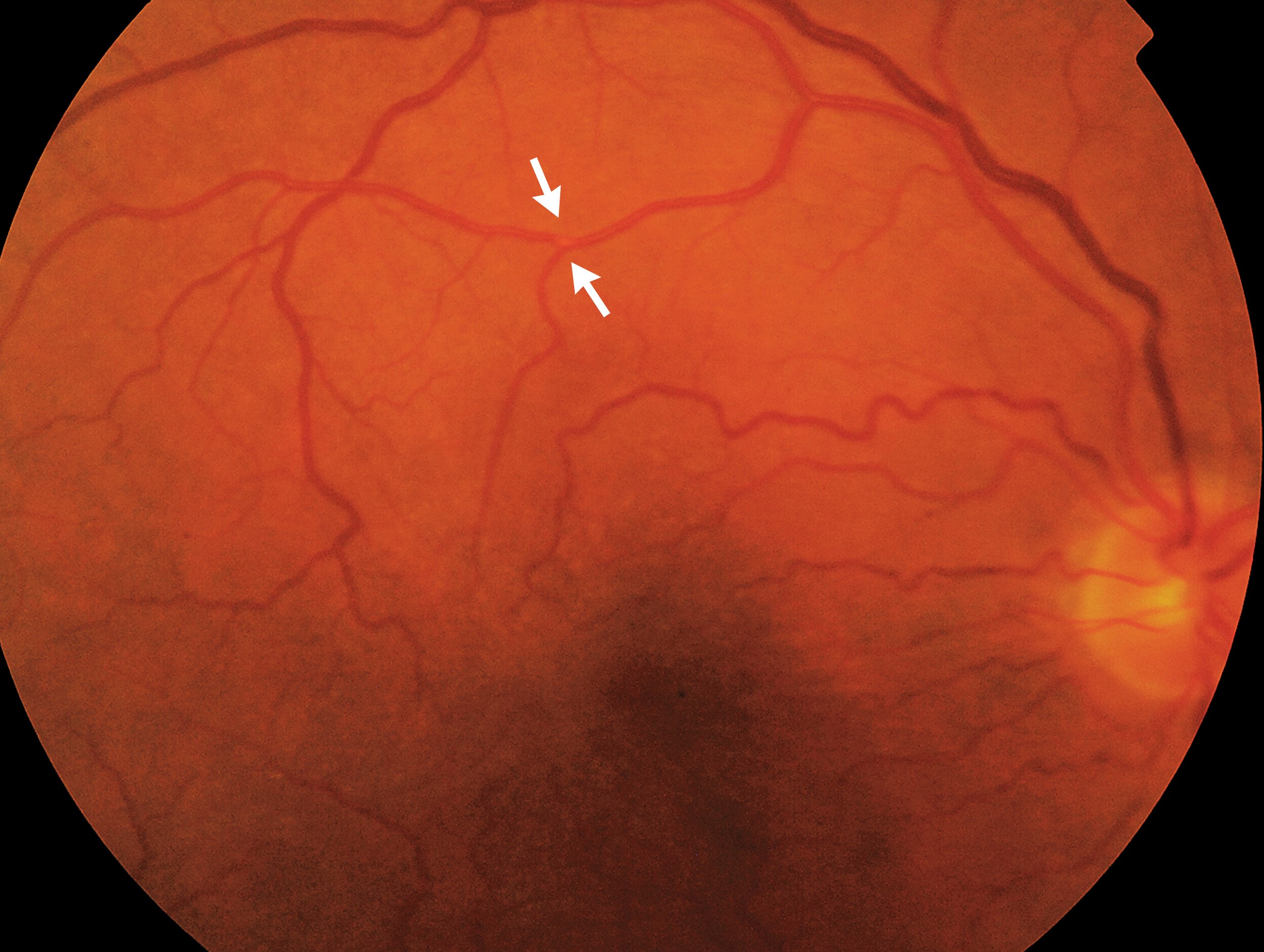
- Hollenhorst plaques, which are bright refractile emboli at arteriolar bifurcations (see Hollenhorst plaque)
- Cotton wool spots, retinal whitening, or a cherry-red spot from a small arterial occlusion
- Venous stasis retinopathy and midperipheral hemorrhages in ocular hypoperfusion
- Optic disc swelling, pallor, or drusen
- Relative afferent pupillary defect
- A carotid bruit and blood pressure in both arms
- Irregular pulse and cardiac murmur
A normal examination does not exclude retinal ischemia.
Investigations
Urgent Assessment
Retinal ischemia is a medical emergency in the same way as cerebral ischemia.
Patients with recent episodes of transient monocular vision loss should be assessed the same day when possible, with vascular imaging and stroke service input.
Investigations include:
- ESR, CRP, and platelet count in patients over 50, to screen for giant cell arteritis
- Carotid imaging with duplex ultrasound, CT angiography, or MR angiography
- ECG and prolonged cardiac rhythm monitoring
- Echocardiography, particularly when a cardiac source is suspected
- Blood glucose, lipids, and full blood count
- Coagulation and thrombophilia testing in young patients without a source
- Brain MRI with diffusion-weighted imaging, which shows silent infarcts in a proportion of patients
Ophthalmic Testing
OCT, fundus photography, and fluorescein angiography help identify retinal ischemia, emboli, and arterial filling delay.
Visual fields and OCT of the optic nerve head help identify optic neuropathy or papilledema.
Management
Vascular Causes
Treatment follows stroke prevention guidelines.
- Antiplatelet therapy, most often aspirin or clopidogrel, started promptly
- Statin therapy and blood pressure control
- Management of diabetes and smoking cessation
- Anticoagulation for atrial fibrillation
- Carotid revascularization for symptomatic high-grade stenosis
In the North American Symptomatic Carotid Endarterectomy Trial, endarterectomy for symptomatic stenosis of 70 to 99 percent reduced the two-year risk of ipsilateral stroke from 26 percent with medical therapy to 9 percent.
The benefit was smaller for moderate stenosis of 50 to 69 percent, and revascularization is most effective when performed early after the event.
Patients with transient monocular blindness had a lower stroke risk than those with hemispheric attacks, but the risk was still significant.
Giant Cell Arteritis
Suspected giant cell arteritis needs high-dose corticosteroids started at once, without waiting for temporal artery biopsy results.
Visual loss from GCA is often permanent and may involve the second eye within days.
Other Causes
Papilledema and demyelinating optic neuropathy are managed by treating the underlying condition.
Retinal migraine is a diagnosis of exclusion and is treated with migraine prophylaxis.
Angle closure is treated with laser iridotomy.
Prognosis
The risk of stroke after retinal TIA is highest in the first days to weeks.
Prompt evaluation and treatment lower that risk considerably.
Patients should understand that a brief episode of visual loss is not harmless, even when vision returns to normal and the eye examination is clear.


Document what you see
Two smartphone imaging tools built for everyday clinic use — one for the slit lamp, one for the fundus.
From Choroida — the team behind this siteReferences
- North American Symptomatic Carotid Endarterectomy Trial Collaborators. Beneficial effect of carotid endarterectomy in symptomatic patients with high-grade carotid stenosis. N Engl J Med. 1991;325:445-453.
- Benavente O, Eliasziw M, Streifler JY, Fox AJ, Barnett HJ, Meldrum H. Prognosis after transient monocular blindness associated with carotid-artery stenosis. N Engl J Med. 2001;345:1084-1090.
- Biousse V, Nahab F, Newman NJ. Management of acute retinal ischemia: follow the guidelines! Ophthalmology. 2018;125:1597-1607.
- Kleindorfer DO, Towfighi A, Chaturvedi S, et al. 2021 guideline for the prevention of stroke in patients with stroke and transient ischemic attack: a guideline from the American Heart Association/American Stroke Association. Stroke. 2021;52:e364-e467.
- Donders RC; Dutch TMB Study Group. Clinical features of transient monocular blindness and the likelihood of atherosclerotic lesions of the internal carotid artery. J Neurol Neurosurg Psychiatry. 2001;71:247-249.
