
Laser pointer maculopathy is a focal foveal injury caused by direct exposure of the eye to a high-power handheld laser.
It occurs most often in children and adolescents who stare at the beam or point it at their own eye, and it can leave a permanent central scotoma.
The blink reflex protects the eye from low-power devices, but many commercially sold pointers exceed their labeled output and can damage the retina in less than a second.
Recognition depends on the history, which patients may not volunteer, and on high-quality OCT.
How Laser Pointer Maculopathy Occurs
Retinal laser injury results from photothermal, photomechanical, or photochemical damage, and the type depends on wavelength, power, and exposure time.
The lens focuses collimated light onto a small area of the fovea, increasing irradiance by several orders of magnitude.
The retinal pigment epithelium absorbs much of the energy, and photoreceptor outer segments are damaged through heat and free radical formation.
Laser products are classified by power.
Class 2 devices (under 1 mW) are considered safe because of the blink reflex, whereas class 3B and class 4 lasers (above 5 mW and above 500 mW, respectively) can injure the retina.
Blue and green handheld lasers sold online often deliver outputs far above their labeled class.
Clinical Presentation
Patients report sudden blurred central vision, a central scotoma, or distortion soon after the exposure.
Some notice an afterimage, a colored spot, or photopsia.
Pain is absent.
Injury may be bilateral when the patient looked at a reflected beam or when a group was exposed.
Visual acuity ranges from normal to severely reduced, depending on the location and depth of the lesion.
Fundus Findings
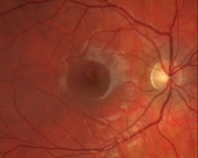
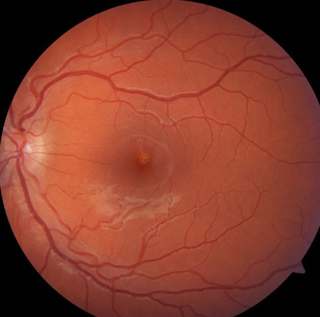
Acute lesions are yellowish-white foveal or parafoveal spots, sometimes with subretinal fluid or hemorrhage.
Multiple lesions or a linear pattern suggest a moving beam.
Later the lesion becomes a pigmented or atrophic scar with a small foveal defect.
OCT
OCT is the most useful test.
- Acute phase: hyperreflective lesion in the outer retina at the fovea, with disruption of the ellipsoid zone and interdigitation zone
- Subacute phase: focal loss of outer retinal layers with a hyporeflective cavity or spike
- Chronic phase: foveal outer retinal defect, sometimes with RPE thickening and atrophy
Autofluorescence often shows a central area of hypoautofluorescence with a ring of hyperautofluorescence.
Fluorescein angiography may show a window defect and, rarely, secondary CNV.
Fundus Explorer Pro
Photograph the retinal findings described here with the phone already in your pocket — 22 D optics and built-in illumination in one handheld unit.
From Choroida — the team behind this siteDifferential Diagnosis
- Solar retinopathy, which shows a similar OCT pattern, and a history of sun-gazing (see solar retinopathy)
- Other photic injuries, such as welding arc and ophthalmoscope injury
- Acute macular neuroretinopathy (see acute macular neuroretinopathy)
- Commotio retinae (see commotio retinae)
- Macular hole
- Vitelliform lesions and other macular dystrophies
- Functional visual loss, which may be suspected in adolescents when the fundus looks normal and OCT is not checked
Laser injury and solar retinopathy can look alike on OCT, and the history is usually what separates them.
Management
There is no proven treatment.
Corticosteroids, hyperbaric oxygen, and other therapies have been tried in isolated reports, and none has been shown to improve outcome.
Practical steps include:
- Obtain a detailed history, including the type of laser, the duration of exposure, and whether others were exposed
- Perform OCT and autofluorescence at baseline and follow-up
- Monitor for secondary CNV in the first months, particularly if there is new distortion
- Provide counseling on prognosis and on avoiding further exposure
- Report the incident to the appropriate authority when a product sold for commercial use is responsible
Adolescents with a laser injury may have behavioral or psychological difficulties, and a supportive approach helps.
Which Lasers Cause Injury
The injury depends on power, wavelength, and exposure time, and not on the appearance of the device.
- Lasers labeled as class 2 or 3R are usually safe when the blink reflex operates, although a determined stare can still overcome it
- Green and blue lasers at several hundred milliwatts can injure the retina in a fraction of a second
- Some devices sold as pointers are functionally class 4 lasers and can also ignite materials and injure the skin
- Blue lasers near 445 nm carry both thermal and photochemical risk, and injuries have been reported after brief exposure
Patients often bring the device to the clinic, which allows the power output to be checked.
Documenting the product, its labeled power, and the circumstances helps with safety reporting and, where relevant, with medicolegal questions.
Prevention
Education is the main preventive measure.
Parents and teachers should be told that laser pointers are not toys, and that high-power devices should not be sold to children.
Ophthalmologists can support regulation of retail products and public warnings about laser eye injuries.
Prognosis
Visual outcome varies.
Many patients improve partly over weeks to months as edema resolves, and some regain near-normal acuity.
Others keep a permanent central scotoma and reduced acuity.
Eyes with large foveal defects or hemorrhage usually do worse.
Patients should be warned that residual scotoma is common even when acuity improves.


Document what you see
Two smartphone imaging tools built for everyday clinic use — one for the slit lamp, one for the fundus.
From Choroida — the team behind this siteReferences
- Mainster MA, Stuck BE, Brown J. Assessment of alleged retinal laser injuries. Arch Ophthalmol. 2004;122:1210-1217.
- Alsulaiman SM, Alrushood AA, Almasaud J, et al. High-power handheld blue laser-induced maculopathy: the results of the King Khaled Eye Specialist Hospital Collaborative Retina Study Group. Ophthalmology. 2014;121:566-572.
- Barkana Y, Belkin M. Laser eye injuries. Surv Ophthalmol. 2000;44:459-478.
- Birtel J, Harmening WM, Krohne TU, Holz FG, Charbel Issa P, Herrmann P. Retinal injury following laser pointer exposure: a systematic review. Dtsch Arztebl Int. 2017;114:831-837.
- Linton E, Walkden A, Steeples LR, et al. Retinal burns from laser pointers: a risk in children with behavioural problems. Eye (Lond). 2019;33:492-504.
