
DISEASE
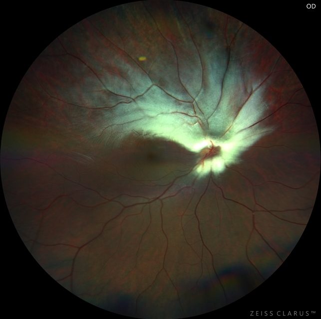
Straatsma syndrome is a rare disease entity characterized by the traditional triad of unilateral myelinated retinal nerve fibers (MNRF), axial myopia, and amblyopia.
The original description of the condition also included strabismus. Variations of the triad include a “reverse Straatsma syndrome ”, in which patients exhibit hyperopia instead of myopia.
Even though nystagmus and strabismus have not been prominently associated with Stratsma syndrome, either may be present as complimentary findings without precluding one from the diagnosis.
Even though Straatsma syndrome has traditionally been considered a unilateral disorder, cases of bilateral Straatsma syndrome and bilateral reverse Straatsma syndrome have also been reported.
Diagnosis
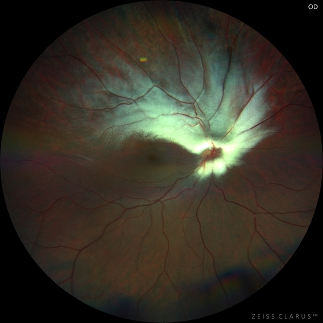
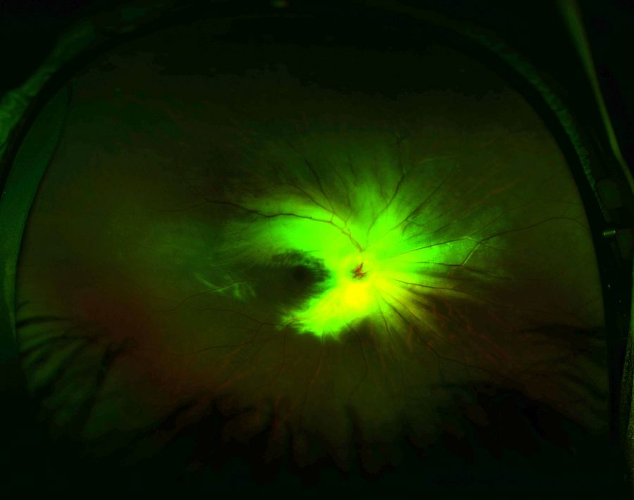
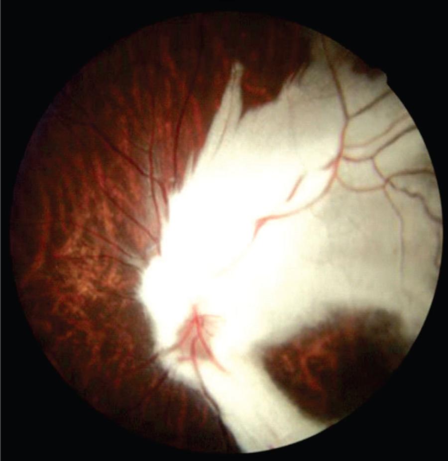
MRNF appears as a white lesion usually contiguous with the optic nerve head. The distribution is along the retinal nerve fiber layer and typically feather-like appearance is noted at the margin.
The MRNF does not cross the horizontal raphe. MRNF may be separate from the optic disc and may be present in the peripheral retina.
Despite some controversy in its diagnostic features, it should be included in the differential diagnosis of leukocoria and must be suspected in the presence of refractive errors with anisometropia.
Fundus Explorer Pro
Photograph the retinal findings described here with the phone already in your pocket — 22 D optics and built-in illumination in one handheld unit.
From Choroida — the team behind this siteMANAGEMENT
The management includes providing proper refractive correction and ruling out systemic associations.
Prognosis
The visual prognosis of amblyopia associated with myelination of retinal nerve fibers and anisometropia is poorer than anisometropic amblyopia without myelination.
It is well known that the former is refractory to occlusive therapy. Despite having a poor prognosis, visual rehabilitation should be attempted.


Document what you see
Two smartphone imaging tools built for everyday clinic use — one for the slit lamp, one for the fundus.
From Choroida — the team behind this siteREFERENCES
- Juhn, A. T., Houston, S. K., & Mehta, S. (2015). BILATERAL STRAATSMA SYNDROME WITH NYSTAGMUS. Retinal Cases & Brief Reports, 9(3), 198–200. doi:10.1097/icb.0000000000000137
- Straatsma BR, Heckenlively JR, Foos RY, Shahinian JK. Myelinated retinal nerve fibers associated with ipsilateral myopia, amblyopia, and strabismus. Am J Ophthalmol. 1979 Sep;88(3 Pt 1):506-10. doi: 10.1016/0002-9394(79)90655-x. PMID: 484678.
- Shenoy R, Bialasiewicz AA, Al Barwani B. Bilateral hypermetropia, myelinated retinal nerve fibers, and amblyopia. Middle East Afr J Ophthalmol 2011;18:65–66
- Levy NS, Ernest JT. Retinal medullated nerve fibers. Arch Ophthalmol 1974;91:330–331
- Congenital optic disk anomalies [Internet]. W.B. Saunders; 2013 [cited 2021 May 19]. Available from: https://www.sciencedirect.com/science/article/pii/B9780702046919000510
- Rosen B, Barry C, Constable IJ. Progression of myelinated retinal nerve fibers. Am J Ophthalmol. 1999 Apr;127(4):471-3. doi: 10.1016/s0002-9394(98)00377-8. PMID: 10218709.
- Quezada-Del Cid NC, Zimmermann-Paiz MA, Ordoñez-Rivas AM, Burgos-Elías VY, Marroquin-Sarti MJ. Straatsma syndrome: Satisfactory amblyopia treatment. Report of two cases. Arch Soc Esp Oftalmol. 2018 Jun;93(6):300-302. English, Spanish. doi: 10.1016/j.oftal.2017.12.010. Epub 2018 Feb 15. PMID: 29398227.
Test yourself
A few questions straight from this article.
-
What is the classic triad of Straatsma syndrome?
Straatsma syndrome is characterised by the traditional triad of unilateral myelinated retinal nerve fibres, axial myopia and amblyopia. -
Which additional feature was part of the original description of Straatsma syndrome?
The original description of the condition also included strabismus alongside the myelinated fibres, myopia and amblyopia. -
What defines the variant known as reverse Straatsma syndrome?
In reverse Straatsma syndrome patients show hyperopia rather than the myopia of the traditional triad. -
What is the typical laterality of Straatsma syndrome?
Straatsma syndrome has traditionally been considered a unilateral disorder, though bilateral cases and bilateral reverse cases have been reported. -
How do myelinated retinal nerve fibres typically appear on fundus examination?
Myelinated fibres appear as a white lesion usually contiguous with the optic nerve head, distributed along the nerve fibre layer with a feather-like margin. -
Which anatomical boundary do myelinated retinal nerve fibres respect?
Myelinated retinal nerve fibres follow the nerve fibre layer and do not cross the horizontal raphe, although they may lie separate from the disc or in the periphery. -
Straatsma syndrome should be included in the differential diagnosis of which clinical sign?
Despite controversy over its diagnostic features, Straatsma syndrome belongs in the differential diagnosis of leukocoria and should be suspected with anisometropic refractive error. -
What does management of Straatsma syndrome consist of?
Management centres on providing proper refractive correction and on ruling out systemic associations, rather than on any retinal intervention. -
How does amblyopia in Straatsma syndrome compare with anisometropic amblyopia without myelination?
Amblyopia associated with myelinated nerve fibres and anisometropia carries a poorer prognosis and is refractory to occlusive therapy, though rehabilitation should still be attempted.
